Add Row
Add Element
Parallel Health World News Logo
update

Parallel Health World

cropper
update
Add Element
  • Home
  • Categories
    • EcoHealth Trends
    • Healing Naturally
    • Age-Defying Diets
    • Supplement Savvy
    • Mind-Body Synergy
    • Finance and Health
    • Biolimitless
    • Tech Hacks
    • Health & Medicine
    • Political
    • BioBuzz
    • Holistic Rehabilitation Techniques
    • Practitioner Insights
    • AI In Healthcare
  • Featured Business Profiles
September 09.2025
1 Minute Read

Act Now: The Window to Fix Your AI in medical image processing Is Closing

Did you know that while more than 80% of hospitals plan to deploy AI in medical image processing by 2025, only a third are confident their systems are truly robust or free from bias? This shocking gap isn't just a statistic—it's a loud wake-up call for everyone from radiologists to hospital CIOs. Right now, we are at a crossroads: act swiftly and fix the cracks in AI algorithms and oversight, or risk compromising both patient care and future innovation. In this comprehensive editorial, we’ll explore why the clock is ticking, the challenges that remain, and why taking decisive steps today will shape the next era of medical imaging.

A Startling Reality: The Current State of AI in Medical Image Processing

The landscape of AI in medical image processing is rapidly transforming, yet lagging behind in critical areas like reliability, transparency, and bias mitigation. While artificial intelligence promises enormous improvements—such as faster diagnostics, optimized treatment planning, and even predictive analytics for diseases like lung cancer or breast cancer—many deployed AI systems still struggle with systemic weaknesses. These include insufficiently diverse imaging data, unexplained neural network decisions, and inconsistencies in regulatory oversight that ultimately place patient outcomes at risk.

Today, leading hospitals and clinics are in the process of integrating AI tools for everything from image segmentation to anomaly detection. However, the rate of actual adoption is not keeping pace with the promises of deep learning and machine learning. As a result, many institutions are caught between the accelerating pressure to modernize and the reality that their AI algorithms are still nascent, often opaque, and sometimes inconsistent across different medical images. The urgency here stems from the possibility that, unless addressed now, these limitations could solidify and create long-term barriers to equitable, safe, and effective healthcare.

AI in medical image processing: concerned doctors and IT specialists reviewing AI results in a modern hospital radiology department, with advanced imaging machines in the background

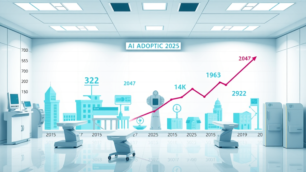
Unveiling the Numbers: AI Adoption in Medical Imaging

"Over 80% of hospitals plan to deploy AI in medical image processing by 2025, yet only 30% have robust, bias-free systems ready."

These figures underscore a dangerous dichotomy in the medical imaging field. As medical imaging tech advances at an unprecedented rate, the groundwork underpinning successful, fair implementation of AI in medical imaging is being laid unevenly. This discrepancy means many health organizations face the risk of deploying AI solutions that could perpetuate existing biases in imaging data, compromise diagnostic accuracy, and impact patient care—especially for underrepresented groups.

Notably, the current momentum among healthcare institutions to implement AI tools stems from the clear benefits AI algorithms and convolutional neural networks promise: scalable diagnostic platforms, more accurate radiological reads, and the ability to handle a deluge of digital medical images. Yet, with so much at stake, the industry must confront the fact that progress in artificial intelligence alone cannot guarantee better patient outcomes without a concurrent commitment to mitigating bias, ensuring data representativeness, and increasing explainability in deep learning systems.

What You'll Learn About AI in Medical Image Processing

  • Why urgency matters: the shrinking window for reliable AI in medical image processing
  • Major obstacles and opportunities shaping AI in medical imaging
  • Expert insights and government perspectives on artificial intelligence in healthcare imaging
  • Actionable steps for institutions, radiologists, and decision-makers

Infographic showing AI adoption rates in healthcare, with overlays of hospitals and radiology equipment for AI in medical image processing

Why the Rush? The Shrinking Window to Fix AI in Medical Image Processing

Decisive action is needed now because the technological evolution in medical imaging is outpacing the careful assessment, standardization, and regulation required to ensure safe deployment of AI systems. As momentum builds—with new learning algorithms and AI tools rolled out at an increasing clip—the window to implement robust, bias-resistant frameworks is narrowing. If stakeholders wait, systemic flaws could become entrenched, eroding both diagnostic accuracy and public trust.

The opportunity to make meaningful course corrections is truly time-sensitive. Investment in better imaging data curation, integration of human eye oversight, and improvement of explainability in AI algorithms needs to keep pace with advances in machine learning. Otherwise, hospitals stand to inherit AI systems that are powerful yet fundamentally limited—putting patient outcomes and even regulatory compliance on the line.

Technological Momentum: Medical Imaging Outpacing Standards

Advanced AI interface analyzing medical scans in a futuristic radiology lab, symbolizing rapid change in AI in medical image processing

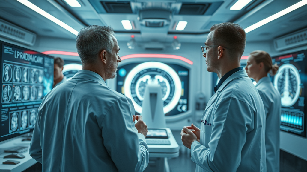
Clinical imaging innovation is accelerating rapidly with widespread use of deep learning, machine learning, and convolutional neural networks for analyzing complex medical images. Algorithms are now capable of identifying early signs of diseases like breast cancer and lung cancer faster than ever, promising a step-change in patient care. However, this technological velocity often surpasses the pace at which ethical, regulatory, and technical standards are updated—another risk factor that demands attention.

For instance, while an AI tool might achieve spectacular diagnostic accuracy in a research setting, its performance can drop dramatically in the real world if imaging data used for training is not diverse enough. This is why technology-driven environments need mechanisms for continuous validation and recalibration—without these, the gap between capability and trustworthiness in medical imaging will only widen.

Systemic Risks: Bias, Error, and Liability in Algorithmic Medical Image Analysis

One of the gravest concerns in deploying AI algorithms for medical image analysis is the risk of ingrained bias—whether in the imaging data used to train neural networks or in the modeling assumptions of the AI system itself. These biases can lead to disparate accuracy rates across demographics, making the role of continuous human supervision and standardized testing indispensable.

Errors in AI systems used for medical imaging introduce unique liability and ethical questions that few institutions are fully equipped to handle. Beyond individual misdiagnoses, the propagation of unchecked bias or error means at-scale harm to entire patient populations. To ensure improved patient outcomes, leaders in healthcare must double down on building transparent, auditable, and well-governed AI in medical solutions before mass adoption is complete.

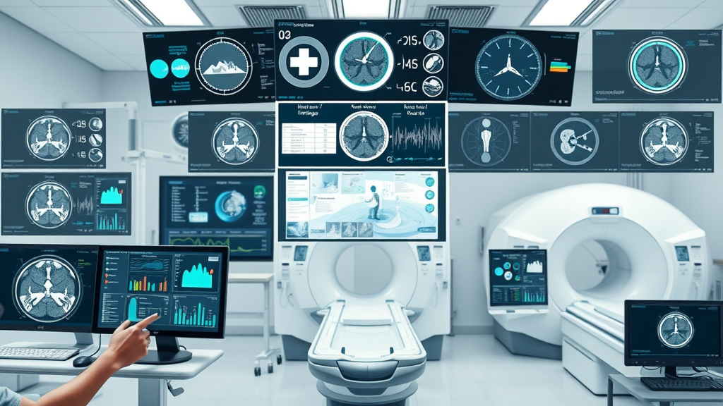
How AI in Medical Image Processing Is Reshaping Healthcare

The introduction of AI in medical imaging is fundamentally altering the future of diagnostics, patient tracking, and care delivery. Using machine learning and deep learning algorithms, these systems can process vast quantities of medical images rapidly, identifying subtle patterns that the human eye might overlook. From reducing turnaround times for critical image reads to helping personalize treatment planning, AI-driven workflows are making real differences—but only when implemented judiciously and ethically.

Particularly, advances in image segmentation, feature extraction, and AI-driven anomaly detection already demonstrate how neural networks and convolutional neural networks can augment radiological interpretation. However, realizing the full promise of AI in medical image processing still hinges on balancing automation with ongoing human oversight and tackling challenges around explainability, generalizability, and equitable training data.

Case Study: Deep Learning Advancements in Breast Cancer Detection

One illustrative example comes from breast cancer screening, where deep learning models are now capable of identifying malignant features on mammograms with accuracy rivaling—or sometimes exceeding—experienced radiologists. Here, AI algorithms trained on vast banks of medical images can spot early lesions, reduce diagnostic subjectivity, and help prioritize follow-up for suspicious findings. Research has shown this can lead to earlier interventions and, in many cases, improve patient outcomes especially for hard-to-detect cases.

Yet, it’s essential to note that these systems often struggle when exposed to image variations outside their training set—for instance, data from different types of scanners, or new population groups. To maximize real-world benefits of AI in medical imaging, models must be continually updated, validated, and overseen by clinical experts to avoid missing rare pathologies or amplifying existing disparities in diagnostic accuracy.

Machine Learning & Imaging Data: Revolutionizing Patient Outcomes

Machine learning runs on the backbone of well-labeled, representative imaging data. When properly harnessed in medical image processing, these algorithms excel at recognizing subtle, complex features invisible to even experienced radiologists. For instance, they detect nuances in lung cancer nodules or microcalcifications in mammography scans, facilitating early signs detection and better treatment planning.

The use of learning algorithms—especially convolutional neural networks—has improved performance in automated image segmentation, organ delineation, and quantification of tumors, directly leading to improved patient outcomes. But this progress also relies on the quality, diversity, and scope of input image data, and highlights the critical need for ongoing data curation and model retraining as clinical scenarios evolve.

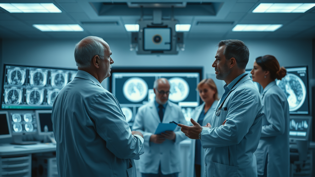
Artificial Intelligence and Human Oversight: The Delicate Balance in Medical Imaging

Radiologist collaborating with an AI-powered computer to analyze a medical scan for AI in medical image processing

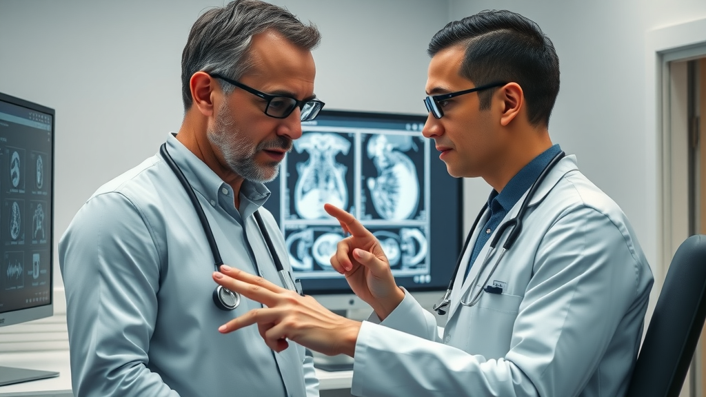
While AI brings computational power and pattern recognition capabilities beyond human reach, their integration into medical image interpretation is never a case for sidelining clinicians. Instead, the next generation of AI in medical image processing is defined by thoughtful collaboration between AI systems and human radiologists, leveraging the strengths of both while mitigating the risk of relying solely on automated outputs.

This human-AI partnership is critical for reducing errors. Human experts catch context-specific subtleties and provide real-time feedback on algorithmic performance, while AI automates the detection of well-characterized patterns, quantifies subtle features, and quickly processes massive image sets. This synergistic approach is central to scalable, high-quality patient care in a rapidly digitizing healthcare environment.

Medical Image Interpretation: What Machines Miss and Humans Catch

Even the most sophisticated artificial intelligence models can stumble on atypical presentations or rare pathologies that aren't well-represented in their training imaging data. Radiologists contribute essential contextual and experiential knowledge, identifying clues that an AI system might miss, such as subtle background abnormalities or non-standard imaging artifacts. The result is a really robust safety net—one that leverages the precision and speed of AI algorithms with the nuanced judgement of the human eye.

Ultimately, the most effective solutions aren’t about replacing radiologists, but augmenting them. This hybrid approach is especially essential for complex diagnoses, uncertain cases, and evolving disease presentations where the context, history, and whole-patient perspective matter as much, if not more, than pure image analysis.

Patient Care Considerations: From Image Analysis to Improved Patient Outcomes

Patient care extends beyond accurate image reads. Integration of AI in medical image processing impacts everything from faster triage and streamlined treatment planning, to reducing unnecessary procedures and ensuring equitable access to leading-edge diagnostics. AI-driven workflows can shorten waiting times, optimally route patients to the right experts, and even provide second-read support—all which directly impact patient outcomes.

But this newfound efficiency must never overshadow the human touch essential to medicine. Empathy, clear communication, and holistic understanding should remain at the center, guiding both the development and deployment of AI tool solutions. Only by prioritizing patient care at every step can AI fulfill its promise as a genuine improvement in healthcare—not just for the technology’s sake, but for people’s lives.

State of the Market: AI Tools in Medical Imaging Today

The market for AI in medical image processing is now home to a growing array of AI tools that claim to automate everything from simple measurements to complex lesion detection. Global investment and VC interest reflect the sector’s transformative potential, but this proliferation also brings a sea of options and little standardization—making selection, integration, and validation difficult for healthcare leaders.

Vendors tout solutions for specific specialties—like AI-driven breast cancer detection, lung cancer screening, or organ segmentation—but not all tools are created equal. Differences in training data scope, regulatory approval (such as FDA clearance), and performance transparency challenge hospitals to separate robust clinical partners from experimental offerings. As the market matures, user-friendly interfaces, integration with existing PACS/EHR, and real-world validation data are quickly emerging as essential markers of reliable AI for medical imaging.

Market Leaders: The AI Tool Landscape

Collage of leading AI medical imaging tools, software dashboards, and MRI equipment for AI in medical image processing

Several companies stand out within the AI tool market, each targeting different modalities and specialties. Leaders offer end-to-end AI platforms capable of handling a variety of medical images—CT, MRI, ultrasound, and digital x-rays—while ensuring interoperability and security of patient data. These solutions are shaped by their ability to demonstrate clear improve patient outcomes, gain regulatory clearance, and offer support for continuous improvement as imaging protocols evolve.

Other challengers take a more focused approach, creating best-in-class solutions for single applications such as image segmentation of brain tumors or early detection in breast cancer screenings. Evaluating these tools requires rigorous side-by-side testing for diagnostic accuracy, usability, integration ease, and transparency of the underlying AI algorithm. Successful deployment depends as much on organizational readiness to adopt and monitor these AI tools as on the technology itself.

Barriers to Broad Adoption in Medical Image Processing

Despite the range of available tools, comprehensive adoption of AI in medical imaging faces persistent obstacles. Core challenges include inconsistent standards for imaging data, a lack of universally accepted protocols for training deep learning systems, and ongoing concerns about how “black box” AI algorithms reach their decisions. Patient privacy and data-sharing constraints complicate the assembly of diverse, high-quality datasets necessary for robust model development and validation.

Additionally, many clinical deployment hurdles remain—from integration with existing radiology workflows to ensuring AI system outputs are interpretable and actionable by human experts. Meeting these challenges will require concerted collaboration between industry, regulators, and medical professionals—and action must be taken now before today’s limitations become tomorrow’s unfixable defects.

Comparison of Leading AI in Medical Image Processing Tools
AI Tool Specialty/Use-Case Strengths Weaknesses Regulatory Status
Al Detect Pro Breast Cancer Screening High sensitivity, fast workflow integration Black box decisions, limited cross-population data FDA cleared
PulmoNet Lung Cancer Nodule Detection Advanced deep learning, multi-modal support Requires large training datasets, explainability issues Pending approval
CardioScan AI Cardiac MRI/CT Analysis Detailed segmentation, clinician dashboard Integration challenges, slow on legacy hardware EU MDR/CE certified
NeuroVision Brain Tumor Localization State-of-art neural networks, intuitive UI Lack of pediatric dataset diversity FDA submitted

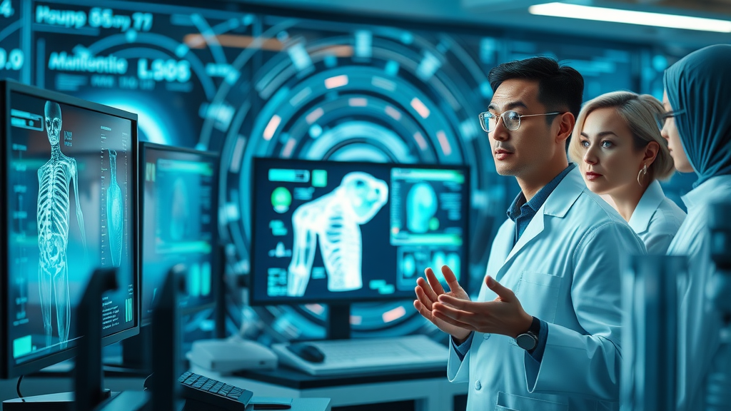
Expert Perspectives on AI in Medical Image Processing

"Human-AI collaboration is the only scalable solution to current bottlenecks in patient care and medical imaging." — Dr. Elaine Park, Radiologist

Expert consensus across radiology, data science, and health informatics highlights the non-negotiable need for collaboration. Leading physicians stress that AI tool outputs must always be interpreted within clinical context, with transparent feedback loops so AI algorithms can be improved and revalidated in real time. Meanwhile, data scientists advocate for more representative and diverse imaging data, and hospital administrators urge for clearer regulatory pathways to allow safe but agile innovation.

Government & Regulatory Viewpoints on Artificial Intelligence in Medical Imaging

Healthcare officials and medical professionals discussing AI policy and regulation for medical image processing

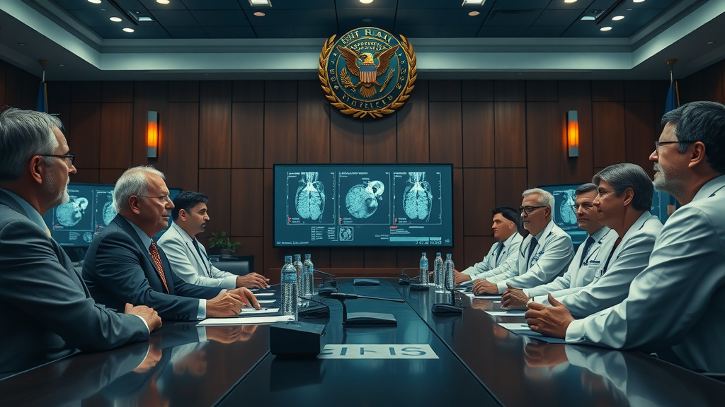
Government agencies and regulators globally are grappling with how to foster safe innovation in AI in medical imaging. The FDA, EMA, and other health bodies are working to define clear pathways for evaluating deep learning models and approving new AI tools for clinical use. A major challenge is keeping regulations responsive to the pace of technological change without compromising on core tenets: safety, equity, and patient data privacy.

Increasingly, policy frameworks emphasize transparency, demands for post-market surveillance, and calls for algorithmic explainability—requiring clear documentation on how AI system decisions are reached. These standards aim to protect patient welfare and public trust, while enabling responsible and ethical scale-up of artificial intelligence in medical imaging.

Key Challenges Facing AI in Medical Image Processing

  1. Data bias in imaging data: Non-representative datasets can result in AI algorithms that underperform for certain populations.
  2. Lack of standardized deep learning protocols: Inconsistent model training impacts reliability and comparability.
  3. Black box algorithms and explainability issues: Clinicians and patients need to understand how AI systems reach medical decisions.
  4. Patient data privacy and ethical considerations: Innovative AI tool development must always uphold the sanctity of patient confidentiality.

Patient Outcomes & The Real-World Impact of Imperfect AI

Concerned patient and doctors reviewing AI analysis on medical imaging results, highlighting the importance of improved, unbiased AI system outcomes.

Inequities and imperfections in AI in medical image processing can have far-reaching consequences on patient care and trust. When AI algorithms misinterpret images due to poor data quality or systemic bias, patients can be subject to misdiagnosis, delayed treatment, or unnecessary procedures—especially in high-stakes contexts like breast cancer screening or lung cancer evaluation.

The potential for improved patient outcomes is immense, but only if all players—technologists, clinicians, and policymakers—move quickly to address known flaws. Redoubled efforts to ensure transparency, accuracy, and ethical development will enable AI in medical imaging to fulfill its promise as a force for good, rather than a source of new risk.

An animated explainer showing how deep learning algorithms analyze medical images, highlighting collaboration between AI and radiologists.

Explore the dual nature of AI in medical image processing—unrivaled opportunity and pressing risk—in this essential perspective video.


People Also Ask: How Is AI Being Used in Medical Imaging?

AI in medical image processing is revolutionizing diagnostics by enabling faster, more accurate interpretation of radiology scans, segmentation of tumors, and pattern recognition in complex imaging data. By integrating deep learning and machine learning, AI tools help radiologists improve patient outcomes and reduce diagnostic errors.

People Also Ask: Can AI Generate Medical Images?

Imaginative AI system generating synthetic medical images for training with amazed researchers observing in a futuristic lab

Yes, AI can generate synthetic medical images for training, research, and managing data scarcity. Generative models and deep learning allow artificial intelligence to create realistic medical image datasets for safer, more robust algorithm development.

People Also Ask: What Is the Role of AI in Healthcare Image?

The role of AI in healthcare imaging spans early disease detection, workflow automation, patient triage, and enhanced image analysis—all of which contribute to better patient care and resource allocation in clinical settings.

People Also Ask: Can AI Do Image Processing?

AI excels at image processing, particularly in medical imaging, where machine learning algorithms automate segmentation, noise reduction, and feature extraction, facilitating more accurate diagnoses and treatment decisions.

Essential Steps Forward: What Needs Fixing in AI for Medical Image Processing

  • Instituting data standardization and reduction of bias
  • Implementing ongoing human oversight
  • Improving regulatory frameworks for artificial intelligence
  • Prioritizing patient outcomes over performance metrics

Visionary group of healthcare professionals, AI engineers, and regulators brainstorming solutions for bias, explainability, and regulation in AI in medical imaging.

FAQs About AI in Medical Image Processing

  • What are the main limitations of AI in medical image processing?
    Primary limitations include data bias, lack of training data diversity, insufficient explainability of how AI algorithms reach conclusions, and challenges with integration into existing clinical workflows. Overcoming these requires collaboration, rigorous validation, and ongoing oversight.
  • How is deep learning different from traditional machine learning in medical imaging?
    Deep learning leverages layered neural networks that automatically extract complex features from imaging data, enabling more nuanced pattern recognition compared to traditional machine learning, which often requires manual feature selection. This allows deep learning to solve harder medical imaging challenges but also demands much larger datasets.
  • Are AI tools FDA approved for clinical use in medical imaging?
    Some AI tools for medical image processing are FDA approved, particularly those with robust clinical validation and safety data. However, many are still under review or in limited use based on specific regulatory pathways. Always check the status and clinical evidence before clinical deployment.
  • How does AI improve patient care in radiology?
    AI in medical imaging boosts patient care by enabling faster, more consistent image reads, early detection of disease, reduction of human error, optimized treatment planning, and helps ensure better allocation of clinical resources. Most importantly, it supports clinicians in making more informed and timely decisions.

Key Takeaways: The Urgency for Robust AI in Medical Image Processing

  • The growth in AI in medical image processing offers immense potential but also introduces urgent challenges.
  • Stakeholders must act now to ensure safe, equitable, and effective implementation.
  • Collaborative regulation, transparency, and patient-centered goals are non-negotiable.

Conclusion: Don’t Let the Window Close on AI in Medical Image Processing

Ignoring the urgency could undermine both patient care and technology’s promise—stakeholders must act decisively today.

AI In Healthcare

0 Views

0 Comments

Write A Comment

*
*
Related Posts All Posts
09.08.2025

Tired of automated image interpretation Holding You Back? Here’s How to Take Control

Over 80% of organizations face bottlenecks in extracting actionable insights due to limitations in automated image interpretation. If you’ve ever struggled with long wait times for results, confusing image analysis outcomes, or feeling like your team isn’t using technology to its fullest, you’re not alone. This comprehensive guide shows you exactly how to overcome common hurdles in automated image interpretation, boost the efficiency of your image analysis pipeline, and implement methods that put you back in control—no matter if you work in healthcare, research, or industry. "Over 80% of organizations face bottlenecks in extracting actionable insights due to limitations in automated image interpretation." Opening Insights on Automated Image Interpretation The explosion of digital images and vast image data across industries has transformed how we process, analyze, and extract meaning. Yet, while the promise of automated image interpretation is enticing—speeding up workflows, reducing manual labor, and providing reproducibility—many organizations find themselves stuck. From the medical image expert trying to detect tumor regions faster, to industrial teams using computer vision for product inspection, bottlenecks persist due to technical, practical, and human challenges. With so many advances in deep learning, neural network tech, and analysis software, taking a critical, informed approach lets you harness the true power of automation without losing oversight or accuracy. This article will demystify automated image analysis from core concepts to practical applications and offer you a roadmap for an efficient, high-quality analysis system. What You'll Learn About Automated Image Interpretation Understanding the foundations of automated image interpretation Key comparison points with manual and hybrid image analysis Step-by-step methods to refine your image analysis pipeline Advanced tools, techniques, and real-world applications Best practices for integrating automation in medical, scientific, and industrial domains Defining Automated Image Interpretation and Its Role in Image Analysis Automated image interpretation is the use of algorithms and artificial intelligence to mimic and often surpass human analysis when extracting information from images. Unlike traditional manual image analysis, which is labor-intensive and subject to human error, automated systems process a wide range of digital images in a fraction of the time. From feature extraction in cell biology to object detection in computer vision tasks, these methods have redefined efficiency. Technologies like machine learning, neural network architectures, and specialized analysis software enable systems to automatically detect features across diverse data sets, including medical images such as MRI or CT scans, industrial inspection images, and field microscopy data sets for scientific discovery. The flow of data has become more streamlined, empowering experts in health care, manufacturing, and research to achieve reproducible, scalable insights that were previously unattainable. Core concepts: automated image, image analysis, image processing Types of data and use cases: medical images, computer vision, field microscopy The Processing Pipeline: How Automated Image Interpretation Works Understanding the detailed description of the processing pipeline is essential for anyone looking to optimize outcomes in automated image interpretation. The typical workflow begins with image processing—including image denoising and normalization—to ensure high-quality, consistent inputs. Next comes segmentation, where the software delineates specific regions, such as a tumor region in a medical image, or objects on a manufacturing line in computer vision. Feature extraction follows, as algorithms measure and quantify relevant attributes, producing reliable image features from huge volumes of data. Finally, classification systems—powered by deep learning and machine learning methods—assign labels or diagnoses, offering insights that drive clinical workflow, industrial decisions, or scientific discovery. Each stage relies on its own set of algorithms, ranging from general-purpose histogram equalization to sophisticated neural networks tailored for specific use cases, ensuring a seamless flow of data from raw imagery to actionable results. Image Processing and Feature Extraction in Automated Image Analysis Image processing is the critical first step for all automated image analysis systems. Here, techniques such as denoising (reducing random noise in digital images), normalization, and restoration prepare the raw data for meaningful analysis. Once the images are prepped, segmentation algorithms separate objects or regions of interest—for example, isolating the tumor region in an oncology dataset or individual cells in field microscopy. Feature extraction is where the heavy lifting happens: quantitative statistics such as texture, shape, or intensity patterns are computed automatically, enabling robust distinction between different tissue types, defects, or sample categories. The automation of feature extraction ensures consistency, reproducibility, and scalability that outpaces manual methods. Instead of slow, subjective measurement, a well-tuned pipeline leverages proven algorithms—like GLCM for texture or U-Net for segmentation—to output a rich set of image features within seconds. Pairing these advancements with a wide range of analysis software, both open-source and enterprise-level, lets teams customize workflows for tasks as diverse as medical image diagnosis, cell lymphoma research, or automated defect detection in industrial quality control. Stages of Automated Image Interpretation Key Tasks Common Algorithms Preprocessing Denoising, normalization Gaussian filter, histogram equalization Segmentation Delineating objects/regions U-Net, thresholding Feature Extraction Quantifying data GLCM, LBP Classification Assigning labels CNN, SVM Deep Learning and Neural Network Techniques in Automated Image Interpretation The surge in deep learning and neural network research has revolutionized the landscape of automated image interpretation. Unlike classic rule-based algorithms, neural networks learn directly from data, automatically detecting intricate patterns and features within complex image data. Convolutional neural networks (CNNs) drive state-of-the-art results in a wide range of medical images, from detecting cancer in radiology to highlighting subtle changes in cell structure for biologists. Deep learning accelerates diagnosis, increases consistency, and frequently matches or surpasses human-level performance in image analysis tasks. These advancements aren’t limited to healthcare. Computer vision applications benefit from neural network models that reliably inspect products, count items, and monitor quality across industrial settings. In field microscopy, deep learning segments and recognizes rare cell types in immense data sets, enabling breakthroughs in diagnostics and life sciences. With a solid processing pipeline, organizations can deploy these technologies to interpret images automatically and improve outcomes, provided that teams also manage data quality, model training, and system oversight to avoid pitfalls such as bias or “black-box” reasoning. Automated Image Interpretation in Practice: Key Applications Automated image interpretation is now a cornerstone in many fields, transforming the speed and accuracy of image analysis. Hospitals deploy AI-powered tools to interpret radiology exams, researchers rely on software to scan microscopy slides for cellular abnormalities, and manufacturers use computer vision to spot flaws before products reach the market. The move toward automation has opened doors for more reliable, efficient decision-making that benefits both end users and industry professionals. By leveraging a robust analysis system—from medical imaging to industrial inspection—organizations handle larger case loads and complex image data without sacrificing quality or consistency. Medical Image Analysis: From Radiology to Oncology "Automated image interpretation has the potential to detect disease markers faster and more consistently than manual analysis." — Dr. Lin, Radiology Expert In the healthcare realm, automated image interpretation has dramatically improved the clinical workflow. Systems powered by deep learning and advanced feature extraction can automatically highlight anomalies in MRI, CT, and histopathology images, often surpassing traditional manual review. This is particularly powerful in oncology, where the quick identification of tumor regions can be life-saving. AI models help radiologists identify abnormalities, segment organs, and even predict disease progression, making diagnostic decisions faster, more consistent, and often more accurate. The integration of automated tools in hospital networks in the United States and beyond ensures scalable, reproducible care across a wide range of medical images. Yet, human oversight remains vital—AI predictions must still be validated by medical experts before clinical decisions are made. Industrial and Scientific Applications: Computer Vision and Field Microscopy Automated image interpretation extends far beyond medicine. In manufacturing, computer vision systems equipped with cameras and neural network algorithms inspect products, identify defects, verify labels, and monitor machinery health—all in real time. This automation improves accuracy, minimizes waste, and scales rapidly to keep up with high-volume production. Scientific research stands to gain, too: automated field microscopy, for instance, enables quick analysis of vast cell or tissue samples, boosting productivity for biologists and clinical laboratories alike. By replacing subjective manual image annotation with standardized, algorithm-driven processes, a wide range of organizations ensure objective data interpretation and make smarter, evidence-backed decisions every day. Advantages and Challenges of Automated Image Interpretation Embracing automated image interpretation comes with clear benefits: workflows become dramatically faster, larger data sets are analyzed with little manual input, and factual, reproducible decisions replace human subjectivity. Whether you’re building a new image analysis system or enhancing a clinical workflow, automation offers scalability, consistency, and often significant cost-effectiveness. However, the journey isn’t without hurdles. Data quality remains a central concern—algorithms trained on limited or biased data can produce misleading or unsafe results. The complexity of deep learning models sometimes leads to “black-box” reasoning, making outcomes difficult to interpret and explain. Maintaining expert oversight and validation steps alongside automation helps strike the vital balance between speed, safety, and accuracy. Ultimately, integrating human experts into the analysis loop ensures the highest level of trust and clinical readiness in both medical and industrial applications. Improved speed and scalability Potential for objectivity and consistency Challenges: Data quality, bias, and interpretability Balancing automation with expert oversight Benefits Challenges Efficiency Data sensitivity Reproducibility Model bias Cost-effectiveness Black-box reasoning Beyond Automation: Achieving High-Quality Image Analysis Results Quality Control in Automated Image Interpretation Regardless of how advanced your analysis software or automation workflow is, maintaining quality control is essential. Benchmarking automated systems against manual workflows helps identify gaps, outliers, or biases in results. For mission-critical fields like medical image analysis—think detecting cell lymphoma or classifying rare diseases—validation with annotated data sets ensures accuracy and reliability. In research and industry, integrating “human-in-the-loop” systems, where experienced analysts review and validate automated outputs, protects against algorithmic mistakes. This practice leverages the strengths of both automation and expert oversight, helping organizations achieve superior, validated results no matter the data set or use case. Benchmarking automated vs. manual workflows Validation with annotated datasets Integrating human-in-the-loop systems Improving Your Automated Image Analysis Pipeline Optimizing your automated image analysis pipeline isn’t a one-time job; it requires ongoing evaluation, collaboration, and innovation. Start by regularly updating training data with new, representative samples to cover a wide range of real-world variability. Continuously benchmark algorithms using both external (public) and internal data to detect drift or degradation in performance over time. Encourage cross-disciplinary collaboration—bringing together data scientists, clinicians, and domain experts—to fine-tune models and ensure output remains relevant for actual decision-making. By nurturing a culture of continuous improvement, you move beyond basic automation to become a leader in extracting maximum value from every digital image your organization encounters. Key Tools and Software for Automated Image Interpretation The landscape of automated image interpretation software is growing rapidly, with robust platforms available for every expertise and budget. Open-source solutions such as ImageJ and CellProfiler provide flexible workflows, extensive plugin libraries, and active community support for both biomedical and general-purpose image analysis. Enterprise platforms like MATLAB and Amira offer advanced analytics, seamless integration with large data sets, and support for scripting custom algorithms tailored to unique industrial or scientific needs. Cloud-based options and AI-powered platforms are making high-end automation accessible to organizations of all sizes, while continuously integrating state-of-the-art advances in deep learning and computer vision. For anyone ready to upgrade their analysis system, understanding the strengths, licensing, and feature sets of these tools is crucial for long-term efficiency and success. Open-source solutions: ImageJ, CellProfiler Enterprise platforms: MATLAB, Amira Emerging cloud-based and AI-powered platforms Watch our introductory video to see how automated image interpretation transforms lab and industrial workflows, featuring real-world cases and easy explanations of the core processing pipeline. Dive into our in-depth video focusing on how deep learning and neural networks are driving breakthroughs in automated image interpretation for medical image analysis, with tangible examples from current hospitals and research labs. People Also Ask: Automated Image Interpretation Is there an AI that can interpret images? Yes, a wide range of AI systems can interpret images through advanced machine learning and deep learning algorithms. These systems—often referred to as automated image analysis tools—can classify, segment, or detect objects and patterns in medical images, satellite imagery, manufacturing data, and more. Examples include convolutional neural networks (CNNs) for medical diagnostics and vision-based inspection platforms for industry. These AI technologies continue to evolve, increasing accessibility and scalability in image analysis workflows around the world. What is automated image analysis? Automated image analysis refers to the process where software interprets digital images without human intervention, usually using artificial intelligence and pattern recognition techniques. This extends from simple measurements (like counting objects) to complex tasks such as diagnosing disease in health care or identifying defects in industrial manufacturing. By leveraging structured processing pipelines—including image processing, segmenting, feature extraction, and automated classification—organizations achieve higher accuracy and efficiency than traditional manual review alone. Can ChatGPT interpret images? As of now, ChatGPT itself is primarily designed for text-based tasks and natural language understanding. However, OpenAI and other platforms are advancing multimodal AI models that combine text and image capabilities, allowing for some level of image interpretation when paired with specialized vision components. For comprehensive automated image interpretation, tools specifically designed for image analysis—utilizing deep learning and computer vision algorithms—are more appropriate and widely used in practice. What are the 7 elements of visual image interpretation? The seven classic elements of visual image interpretation include: shape, size, pattern, tone or color, texture, shadow, and association. These features guide both manual and automated interpretation by providing quantitative and qualitative clues to identify, segment, and classify objects across a variety of digital images, such as medical diagnostics, satellite imagery, or material science samples. FAQs: Automated Image Interpretation What are typical data requirements for automated image interpretation? Robust data requirements usually include well-annotated image data sets that cover all classes of interest, consistent imaging conditions, and high-resolution images where possible. High-quality input ensures algorithms learn the right patterns and generalize well for real-world cases. How do you evaluate the accuracy of automated image analysis? Accuracy is measured by comparing automated results to ground truth annotations using metrics like precision, recall, F1 score, and overall accuracy percentage. Cross-validation on external data and expert review are also crucial for validating the system. Which industries benefit most from automated image? Key benefitting industries include healthcare (radiology, pathology, cell lymphoma detection), manufacturing (quality control via computer vision), life sciences (field microscopy, cell counting), geospatial analysis, and security applications. Can human experts override AI interpretations? Yes, especially in critical applications like medical diagnostics or industrial safety inspections. Many analysis systems include a “human-in-the-loop” design where experts review, validate, or override AI-derived results for maximum accuracy and trust. Is automated image interpretation safe for clinical use? When validated on diverse, well-annotated datasets and supervised by experts, automated image interpretation tools are safe for clinical decision support. Regulatory agencies often require extensive testing and ongoing validation before adoption in clinical workflow. Key Takeaways: Mastering Automated Image Interpretation Automated image interpretation offers transformative efficiency and scalability Success depends on understanding algorithms, validation, and application context Combining automation with expert insight yields the best analytical outcomes Conclusion: Take Charge of Your Automated Image Interpretation By understanding the technology, challenges, and best practices, you can confidently implement automated image interpretation to achieve fast, high-quality results without losing expert control over your workflow.

09.08.2025

Unveil the Secret of neural networks for medical imaging for Breakthrough Accuracy

Startling Fact: Did you know that more than 90% of radiologists report improved diagnostic accuracy thanks to neural networks for medical imaging? These advances are rapidly shrinking the gap between human expertise and artificial intelligence, transforming radiology and patient care as we know it. Let’s dive deep into how these sophisticated deep learning models are setting new standards in medical image analysis—pushing the boundaries of accuracy, speed, and reliability. Opening Insights: The Surprising Impact of Neural Networks for Medical Imaging Neural networks, specifically deep neural networks and convolutional neural networks (CNNs), have taken medical imaging to a revolutionary level. Using advanced deep learning, artificial intelligence can now scan, interpret, and compare complex data from MRI images, CT scans, and X-rays with an accuracy that rivals and often exceeds human performance. The widespread adoption of these neural networks for medical imaging has shifted diagnostic protocols in radiology departments globally, impacting everything from workflow management to patient outcomes. This technology’s power lies in its ability to rapidly process vast quantities of medical data, learning to identify subtle patterns and anomalies. For instance, neural networks excel in image segmentation and image classification, making it possible to spot tumors, microfractures, and rare diseases quickly and reliably. With the addition of transfer learning, these models get even smarter—leveraging knowledge from vast image libraries to boost performance in new domains or limited-data scenarios. As more clinicians witness AI’s tangible results, the case for integrating deep learning into medical image analysis is stronger than ever. “More than 90% of radiologists say that artificial intelligence powered by neural networks for medical imaging has improved diagnostic accuracy.” — Journal of Medical Imaging What You'll Learn About Neural Networks for Medical Imaging The fundamentals of neural networks for medical imaging The latest advances in deep learning and convolutional neural network (CNN) architectures How transfer learning improves medical image classification Expert viewpoints on the superiority of artificial intelligence in healthcare Emerging applications in image segmentation and analysis The Evolution of Neural Networks for Medical Imaging From Artificial Intelligence to Deep Neural Network Breakthroughs The journey of neural networks for medical imaging began with the broader field of artificial intelligence in the late 20th century. Early efforts relied on simple machine learning models that required explicit programming and could process only limited features of medical images. However, the real breakthrough arrived with the advent of deep neural networks, especially deep learning models capable of learning from vast datasets. These networks became adept at pattern recognition, making them ideal for sophisticated image analysis in radiology and pathology. Deep learning, powered primarily by neural network innovations, allowed for multi-layered data extraction. These advances positioned AI as a robust force for medical image analysis by automating feature identification, reducing diagnostic errors, and increasing efficiency in diagnostic imaging. Today’s deep neural networks can analyze CT images, MRI images, and mammograms with remarkable precision. Their progression is marked not only by technological innovation but by a growing acceptance among medical professionals who see artificial intelligence as a strategic partner in delivering patient-centric care. Key Milestones: Transfer Learning & Convolutional Neural Networks One of the defining milestones in this field has been the implementation of transfer learning and convolutional neural network (CNN) architectures. Transfer learning, which leverages pre-trained models, enables a deep neural network trained on one set of medical data to rapidly adapt to new types of scans or rare conditions, minimizing the need for massive labeled datasets. This is especially vital for medical image classification in diseases like breast cancer and rare abnormalities where data is scarce. Convolutional neural networks, designed to mimic the visual cortex of the human brain, specialize in extracting hierarchical features from medical images. These CNN architectures have become the backbone of modern image segmentation, image classification, and anomaly detection systems in medical image analysis. The result is a dramatic improvement in both speed and accuracy, setting new benchmarks for medical imaging. Why Neural Networks for Medical Imaging Outperform Conventional Approaches Superior Diagnostic Image Analysis and Classification What truly sets neural networks for medical imaging apart is their superior capacity for image classification and analysis. Traditional diagnostic methods heavily depend on human expertise, which—while formidable—can be subjective, labor-intensive, and limited by cognitive fatigue. In contrast, deep learning models, especially convolutional neural networks, work tirelessly around the clock, digesting massive volumes of CT images and MRI images with unwavering focus. These models learn to identify intricate features—such as tissue patterns, lesions, or microcalcifications—with consistently high accuracy. In clinical studies, deep neural networks have matched or even outperformed radiologists in tasks like identifying early-stage breast cancer and classifying brain tumors from CT images. This automation not only reduces the margin for human error but also accelerates the workflow, ensuring critical conditions are detected faster and with fewer false negatives or positives. Image Segmentation and Deep Neural Networks for Precision Image segmentation—the process of delineating distinct structures within medical images—has been revolutionized by convolutional neural networks. Unlike earlier manual or semi-automated techniques, deep neural networks can quickly differentiate and label tissues, organs, or anomalies with remarkable detail and consistency. This level of precision is crucial for complex procedures such as surgical planning, tumor monitoring, and multi-modal image analysis. The adaptability of CNN architectures enables them to tackle challenging scenarios, including overlapping structures and poor image quality, that often confound traditional algorithms. Deep learning models are now routinely applied for segmentation in CT scans, MRI images, and even ultrasound data, translating directly to better patient management and more informed clinical decisions. “The accuracy of deep learning models in medical image analysis often matches or exceeds human expertise.” — Leading AI Researcher Critical Components: Deep Learning, Convolutional Neural Networks, and Machine Learning How Deep Neural Networks Transform Medical Image Classification The core transformative agent in today’s radiology labs is deep neural network technology. By structuring models in multiple layers—where each layer identifies increasingly abstract features—deep neural networks turn blurry medical data into actionable clinical insights. This multilayer approach is particularly effective for medical image classification tasks, such as labeling chest X-rays for pneumonia or distinguishing between benign and malignant lesions in breast cancer screening. Convolutional neural network architectures add another dimension by automating feature extraction from raw data. This eliminates manual intervention and paves the way for highly scalable medical image analysis pipelines. Through machine learning, these models are continually retrained and improved, easily adapting to the introduction of new diseases or imaging technology, hence futureproofing radiological practices. Unlocking the Power of Data Augmentation & CNN Architectures A significant factor in the reliability of neural networks for medical imaging is the practice of data augmentation. By artificially expanding existing datasets with rotated, flipped, or modified medical images, data augmentation helps deep neural networks learn robustly from limited or imbalanced data—a common hurdle in healthcare. This, paired with advanced CNN architectures, empowers models to thrive where traditional algorithms may falter. Adaptive CNN architectures go further by automatically calibrating filters and layers, optimizing for varying imaging modalities such as MRI, CT, or X-ray. These enhancements bolster the model’s capacity to generalize across different patient demographics, scan settings, and even hospital equipment. Ultimately, this adaptability translates into real-world effectiveness, driving the evolution of automated medical image analysis. Conventional Algorithms vs Neural Networks for Medical Imaging Method Diagnostic Accuracy Processing Time Adaptability Traditional Algorithms Moderate Slow to Moderate Low (Manual Feature Extraction) Deep Learning/Neural Networks High to Very High Rapid (Real-Time Possible) High (Automatic, Learns New Data) Transfer Learning in Neural Networks for Medical Imaging: A Revolutionary Approach Transfer Learning Applied in Radiology and Beyond Transfer learning is swiftly re-shaping neural networks for medical imaging. By using models initially trained on large, general datasets, transfer learning allows for rapid deployment into specialized fields—like pediatric radiology or rare cancer detection—where labeled data may be scarce. This revolutionary strategy not only cuts down the time and resources needed for model development but also boosts performance for novel or uncommon use cases. Radiology departments have seen particular success, where transfer learning empowers convolutional neural networks to recognize nuances in CT images or adapt to hospital-specific imaging protocols. This cross-domain intelligence enhances diagnostic consistency and allows for immediate upskilling of models in response to new disease outbreaks or novel imaging technologies, a crucial advantage in times of health crises. Real-World Applications: Neural Networks in Medical Imaging Today Detection of Breast Cancer with Neural Networks The detection of breast cancer through medical imaging stands as one of the most celebrated triumphs of neural network technology. Deep convolutional neural networks efficiently scan mammograms for anomalies, microcalcifications, and irregular tissue patterns. These models deliver results at tremendous speed, enhancing both early detection and long-term patient survival rates. Recent advancements enable neural networks to not just highlight potential areas of concern but also provide confidence metrics that help radiologists focus on the highest-risk findings. In practical clinical settings, the ability of AI to sift through extensive datasets, reduce false positives, and adapt instantly to new imaging modalities has led to a noticeable reduction in missed diagnoses. As a result, patients and doctors alike now count on these systems as indispensable decision-making tools in breast cancer care. Neural Networks for Medical Image Segmentation and Analysis Segmentation is essential in multiple stages of healthcare, from targeting radiation therapy to tracking organ changes over time. Neural networks, particularly those based on CNN architectures, shine in segmenting high-resolution medical images, efficiently separating tumor tissues from healthy organs or outlining the boundaries of complex bone structures. These models excel even in conditions where contrast is low or overlapping tissues are present—situations that often challenge conventional techniques. Advanced segmentation capabilities brought by deep neural networks are now fundamental for fields like oncology, orthopedics, and cardiology. They enable more precise surgeries, personalized treatment plans, and accurate disease progression monitoring, giving medical professionals a new lens through which to see and treat their patients. AI and Deep Learning in Image Classification for Rare Disease Detection Rare disease diagnosis can stump even the most experienced clinicians due to the small number of training examples and often subtle imaging signatures. Neural networks for medical imaging address this challenge by employing transfer learning, data augmentation, and sophisticated image classification strategies to recognize anomalies that otherwise might go unnoticed. Deep learning models, equipped with adaptive convolutional layers and a well-structured connected layer, can autonomously flag at-risk patients, escalating cases for human review before symptoms worsen. This autonomous triage system is helping hospitals consistently deliver faster, more accurate, and life-saving care in the hunt for elusive, rare diseases. Challenges and Ethical Considerations in Neural Networks for Medical Imaging Algorithm Transparency, Bias, and Clinical Acceptance Despite their transformative impact, neural networks for medical imaging face real-world challenges. Algorithm transparency—or the lack thereof—remains a major roadblock. Many deep learning models operate as “black boxes,” leaving radiologists and patients uncertain about how key decisions are made. This opacity can undermine trust, especially when critical medical decisions are involved. Equally pressing is the risk of bias, as neural networks trained on imbalanced datasets may inadvertently propagate health disparities—missing disease patterns common in underrepresented groups. Gaining broad clinical acceptance requires ongoing education and the development of explainable AI techniques that allow healthcare professionals to understand and trust deep learning outputs. Balancing Data Privacy with Powerful Deep Learning Capabilities Protecting patient privacy while leveraging powerful AI models is perhaps the most delicate balancing act facing medical imaging today. Large datasets required for training deep neural networks are often rich in sensitive personal information. Ensuring compliance with privacy regulations like HIPAA while enabling the sharing and aggregation of medical data is essential for continuous progress. Techniques such as federated learning and anonymization are rapidly emerging, ensuring that neural networks for medical imaging can be trained robustly without exposing individual identities. Ultimately, ethical stewardship and transparency must go hand-in-hand with technological advancement as the use of artificial intelligence expands in healthcare. The Future: Next-Generation Neural Networks for Medical Imaging Emerging Trends: Explainable AI, Advanced CNN Architectures, and Beyond The next era for neural networks and deep learning in medical imaging is marked by innovations in explainable AI and increasingly advanced convolutional neural network architectures. Explainable AI seeks to open the “black box,” offering logical justifications for each diagnosis or image classification, bolstering both accountability and confidence among medical professionals. New wave CNN architectures, including attention mechanisms, graph neural networks, and unsupervised learning algorithms, are pushing the accuracy, efficiency, and flexibility of models to all new heights. These advancements promise precision medicine—where diagnostics and treatments are uniquely tailored to each patient and supported by a transparent, trustworthy AI companion. Expectations for Transfer Learning and Deep Neural Networks in Healthcare Looking ahead, transfer learning and deep neural networks will remain at the heart of medical advances. As imaging datasets grow, and models learn from ever-diverse data, the precision and adaptability of AI tools will become even more pronounced. The integration of neural networks into electronic health records, real-time operating rooms, and telemedicine will drive global health equity and ensure rapid deployment of life-saving diagnostics anywhere in the world. Personalized, data-driven care powered by neural networks for medical imaging is poised to become standard, not exception. As computational power soars and ethical frameworks mature, the full promise of artificial intelligence in medicine comes into clearer focus—one image at a time. “As neural networks for medical imaging evolve, personalized diagnostics become not just possible, but inevitable.” — Healthcare Futurist People Also Ask: Neural Networks for Medical Imaging What are neural networks used in medical image processing? Neural networks for medical imaging are primarily employed for tasks such as image segmentation, classification, and anomaly detection, enabling the rapid identification of diseases and abnormalities in X-rays, MRIs, CT scans, and more. Which neural network is best for medical image classification? Convolutional Neural Networks (CNNs) are widely recognized as the most effective for medical image classification due to their ability to automatically extract relevant features from complex medical images. What neural network is used in radiology? Radiology leverages deep convolutional neural networks along with transfer learning to analyze large volumes of radiological images with high precision and speed. How are neural networks used in healthcare? Neural networks are employed across the healthcare sector for predictive analytics, diagnostic imaging, patient risk scoring, and personalized treatment recommendations, expanding the frontiers of artificial intelligence in medicine. Expert Opinions: The Transformational Potential of Neural Networks for Medical Imaging “Neural networks are not just tools—they’re partners in diagnosis.” — Renowned Radiologist Lists: Key Benefits of Neural Networks for Medical Imaging High diagnostic accuracy and efficiency Reduction in human error Ability to handle complex and large datasets Rapid adaption to new imaging modalities via transfer learning Enhanced support for radiologists and medical professionals FAQs: Neural Networks for Medical Imaging How do neural networks improve image analysis speed in medical imaging? Neural networks greatly improve image analysis speed by utilizing parallel processing and automated feature extraction. Deep learning models, particularly convolutional neural networks, can process thousands of medical images rapidly, reducing turnaround time for diagnostic results. As a result, clinicians receive critical insights sooner, which can be pivotal in emergencies or high-throughput settings. This efficiency means faster, more accurate care for patients and less backlog for busy imaging departments. Are there risks or drawbacks in using neural networks for medical diagnosis? While neural networks offer impressive accuracy, they are not without risks. The “black box” nature of deep learning can make it challenging to interpret and explain decisions, leading to hesitation among clinicians and patients. There is also the potential for algorithmic bias if models are trained on unrepresentative data, possibly resulting in health disparities. Addressing these challenges requires ongoing research into explainable AI, careful training dataset selection, and continual oversight by medical experts. Can neural networks for medical imaging work with limited datasets? Yes, through methods such as data augmentation and transfer learning, neural networks can operate effectively even when labeled medical datasets are limited. Data augmentation artificially expands training data, while transfer learning adapts pre-trained models to new, smaller datasets. These approaches allow AI-driven medical imaging solutions to be implemented in rare disease detection, pediatric diagnostics, or resource-limited settings without compromising reliability. Key Takeaways: Author’s Reflections on Neural Networks for Medical Imaging Integration of neural networks for medical imaging marks a paradigm shift in diagnostic medicine. Ethical challenges must be met with transparency and rigorous oversight. The growing accuracy and efficiency of deep neural networks promise a future of precision care. Conclusion: Embracing Neural Networks for Medical Imaging as the Future of Diagnosis Neural networks for medical imaging represent not just an upgrade in technology, but a transformation in patient care—where diagnosis is faster, more accurate, and increasingly equitable. “Adopting neural networks for medical imaging is not just about technology—it’s about saving lives through smarter medicine.” Tables: Comparison of Neural Network Architectures for Medical Imaging CNN vs Deep Neural Network vs Traditional Algorithms in Medical Imaging Architecture Best For Accuracy Interpretability Scalability Convolutional Neural Network (CNN) Image classification and segmentation High Moderate Excellent Deep Neural Network (DNN) Pattern recognition, feature extraction Very High Low Very Good Traditional Algorithms Rule-based diagnostics Moderate High Limited Lists: Innovative Tools Empowered by Neural Networks for Medical Imaging Automated tumor detection platforms Real-time anomaly detection systems Advanced image segmentation suites Watch: How Neural Networks are Shaping Medical Imaging — explainer video highlighting the transformation in radiological diagnostics, key visualizations, and expert interviews. Discover real-world examples: Deep Learning in Action presents medical imaging case studies where deep learning and neural network techniques directly accelerated diagnosis and improved patient outcomes.

09.05.2025

How to Solve medical image segmentation AI Without Common Frustrations

Startling Statistic: Did you know nearly 30% of AI-driven segmentation errors in clinical settings arise from overlooked training data biases? While mainstream discussions often celebrate breakthroughs in medical image segmentation AI, they seldom spotlight the hidden pitfalls that can derail even the most sophisticated deep learning approaches. In this opinion-based guide, we unravel the real frustrations behind segmentation tasks—and show you how to solve them with insight and innovation. Medical Image Segmentation AI: Breaking Down the Barriers with Unconventional Insights At the frontlines of advanced healthcare, medical image segmentation AI is rapidly changing how doctors and researchers analyze, diagnose, and plan treatments. Yet, despite the promises of deep learning and the power of modern artificial intelligence, the journey toward flawless image segmentation is filled with unexpected challenges. From inconsistent model performance to biases lurking in training data, practitioners routinely encounter hurdles that slow or even sabotage clinical adoption. This section lays out fresh perspectives and actionable insights to help you move beyond hype, equipping you with a more realistic—and more effective—approach to solving the core issues in medical image segmentation tasks. By uncovering surprising statistics and sharing under-the-radar pitfalls, we'll look at how successful segmentation models aren’t just the result of technological wizardry. Instead, they're products of smart workflow design, critical skepticism, and interdisciplinary teamwork. With practical solutions and a shift in mindset, you can sidestep frustration and develop solutions that truly deliver in demanding medical contexts. Startling Facts: The State of Medical Image Segmentation and Artificial Intelligence The surge in medical image segmentation AI is undeniable, yet beneath this momentum lies a landscape riddled with issues that rarely make headlines. For instance, large-scale research indicates that a significant proportion of model failures—sometimes as high as 30%—are due not to the complexity of medical images, but to invisible issues in training data. Biases such as underrepresented disease types or imaging artifacts can mislead even the most advanced deep learning models, undermining both diagnostic accuracy and practitioner trust. This unexpected source of error highlights the need to re-evaluate our approaches to model training, validation, and deployment. "Nearly 30% of AI-driven segmentation errors in clinical settings arise from overlooked training data biases—an issue rarely spotlighted in mainstream medical imaging discussions." What You'll Learn from This Opinion Piece on Medical Image Segmentation AI Why medical image segmentation AI is more challenging than most believe Practical ways to overcome deep learning pitfalls in image segmentation tasks Personal insights on balancing technological optimism with skepticism How to judge segmentation model performance on medical images realistically Recommended segmentation methods that avoid common frustrations Understanding Medical Image Segmentation AI: The Fundamentals To tackle the pervasive issues in medical image segmentation AI, it’s crucial to first establish a solid foundation. At its core, medical image segmentation involves dividing a medical image (such as a CT scan or MRI) into distinct regions corresponding to different anatomical structures or pathological areas. This process is vital for a wide array of applications, from identifying tumors to planning surgeries, and is powered by advances in both artificial intelligence and computer vision. Success in this space requires an awareness of both traditional and cutting-edge segmentation methods. While deep learning has revolutionized segmentation tasks, traditional techniques such as thresholding, edge detection, and region growing remain relevant—especially when deep learning models hit roadblocks, such as limited or biased training data. Recognizing the strengths and weaknesses of each approach and knowing when to deploy them is a skill set that separates reliable solutions from those plagued with frustration and unreliability. Defining Medical Image Segmentation and Its Role in Artificial Intelligence Medical image segmentation refers to the precise division of medical images into regions or segments that represent different tissues, organs, or pathological areas. This task plays a foundational role in artificial intelligence-powered image analysis. For example, segmenting tumors, organs, or blood vessels in CT and MRI scans is crucial for both diagnosis and treatment planning. Effective segmentation enhances the clarity of input images, allowing AI algorithms to focus on meaningful structures—improving automation and reducing manual annotation in clinical settings. Artificial intelligence—particularly deep learning approaches such as convolutional neural networks (CNNs)—has taken center stage in automating image segmentation tasks. AI models are trained on extensive datasets of annotated medical images, learning to distinguish between normal and abnormal tissue, or to identify subtle changes not easily recognized by human experts. But this reliance on training data and the inherent complexity of biomedical image segmentation means that even minor data inconsistencies can disrupt model performance. This raises the stakes for careful data curation and critical evaluation of segmentation models in practical, real-world settings. Core Segmentation Methods: From Deep Learning to Traditional Approaches Segmentation methods in medical imaging range from classic algorithms—like thresholding and region growing—to modern deep learning models. Traditional techniques typically rely on low-level pixel information, which makes them relatively interpretable but potentially less adaptable to complex variations in biomedical images. In contrast, deep learning models (such as U-Net and Mask R-CNN) learn features directly from data and can capture more intricate patterns. However, they are often “black boxes,” making it difficult to explain segmentation results and diagnose failure modes. Choosing the right segmentation method depends heavily on clinical requirements, the diversity of available training data, and the need for model interpretability. For rare disease segmentation or when datasets are limited, hybrid approaches that combine domain knowledge with machine learning are gaining traction. These methods help balance the flexibility of AI with the reliability of tried-and-true image analysis techniques—critical for ensuring reliable outcomes in the clinical setting. Comparison of Segmentation Models Used in Medical Image Segmentation AI Segmentation Model Type Strengths Weaknesses Thresholding / Region Growing Traditional Simple, interpretable, minimal data requirements Struggles with noise, poor at handling complex structures Active Contour (Snake) Traditional Good for smooth boundaries, interactive adjustment Sensitive to initialization, limited automation U-Net (CNN) Deep Learning High accuracy, robust for biomedical image segmentation, scalable Requires large annotated datasets, less interpretable Mask R-CNN Deep Learning Multi-object segmentation, flexible for varied input image types Computationally intensive, can overfit limited data Hybrid Models (AI+Rules) Hybrid Balances AI learning with domain heuristics, improved interpretability Requires multidisciplinary expertise to implement How Computer Vision Powers Medical Image Segmentation Tasks Computer vision forms the backbone of automated medical image segmentation. Using mathematical and statistical techniques, computer vision enables AI systems to extract meaningful patterns from complex input images—ranging from CT scans to ultrasound images and beyond. At the core, these techniques empower neural network and deep learning models to recognize minute differences in tissue, shape, and texture that may go undetected through manual review. The contribution of computer vision extends beyond pattern recognition. By facilitating semantic segmentation—where every pixel is classified into a relevant category—it streamlines image analysis, enhances diagnostic workflows, and supports vital clinical decisions. This synergy between AI and computer vision is ushering in new standards for speed, accuracy, and reliability in clinical diagnostic settings, but it also presents unique challenges that require cross-disciplinary expertise and ongoing scrutiny. Common Frustrations in Medical Image Segmentation AI and Their Root Causes Many practitioners approach medical image segmentation AI anticipating rapid, transformative benefits. Instead, they're often met with frustrating setbacks that range from wildly inconsistent segmentation results to the persistent failure of deep learning models on real-world datasets. These difficulties reflect deeper, structural issues within the field—such as the absence of high-quality, representative training data, the inadequacy of existing evaluation metrics, and the lack of transparency in segmentation models. Understanding these sources of frustration is the first step toward building robust and reliable solutions for real-world clinical use. This section dives into the tangible root causes—why segmentation performance often falls short of expectations, why training data is both a necessity and a liability, and how semantic segmentation sometimes amplifies rather than eliminates errors. By addressing these points head-on, you’ll gain a realistic perspective—and the tools—for overcoming the challenges unique to medical image segmentation tasks. Segmentation Performance: Why Results Frustrate AI Practitioners Segmentation performance is where the promises and perils of medical image segmentation AI become especially apparent. Practitioners often find that even highly-touted segmentation models underperform on new or unseen datasets. Common causes include a lack of generalizability, overfitting to training data from a single institution, and poor quality control in data labeling. These problems are compounded in sensitive clinical environments, where inconsistent or inaccurate segmentation results can delay or jeopardize patient care. The fact that no single segmentation model performs optimally across all segmentation tasks highlights a crucial limitation: model evaluation must move beyond artificial benchmarks to reflect real-world complexity. Diverse test datasets, rigorous cross-institutional validation, and ongoing clinician feedback are needed to ensure model performance translates into practical clinical utility. Only by acknowledging these nuances can AI practitioners move beyond surface-level solutions and establish reliable standards for medical image analysis. The Training Data Dilemma in Medical Images Training data is the bedrock on which all deep learning and computer vision systems are built; yet, this resource is notoriously difficult to get right in medical imaging. High-quality annotated medical images are expensive and time-consuming to produce. Biases can creep in through overrepresentation of common cases or exclusion of rare diseases and specific patient populations, leading to skewed model performance. Furthermore, privacy regulations and fragmented data sources pose additional barriers to compiling diverse training sets. When training data does not reflect the full complexity of clinical reality, deep learning models may excel on paper but fail in the real world. The challenge for practitioners is to continually expand, refine, and audit their datasets—incorporating ongoing feedback from both machine learning experts and frontline clinicians. Rigorous attention to dataset construction and curation is as important as algorithm selection for trustworthy image segmentation results. Semantic Segmentation Pitfalls: Where Deep Learning Fails Semantic segmentation—where each pixel in a medical image is assigned to a meaningful class—remains a central goal of AI-powered image analysis. However, deep learning models used for semantic segmentation are vulnerable to multiple failure points. These include subtle but critical annotation errors, generalized domain shift problems (when training data differs from deployment scenarios), and a lack of model interpretability, which can mask systematic errors. These vulnerabilities mean that when segmentation fails in the clinical setting, it can erode trust among medical professionals and jeopardize patient outcomes. To minimize such frustrations, leading practitioners recommend rigorous benchmarking across different segmentation tasks, developing explainable segmentation models, and incorporating human-in-the-loop feedback mechanisms. In the world of medical imaging, transparency, interpretability, and collaboration are not optional—they’re essential safeguards. Opinion: Why Medical Image Segmentation AI Deserves a Nuanced Approach "True progress in medical image segmentation will not come from bigger models, but from smarter segmentation methods and honest conversations about limitations." The race to build “state-of-the-art” models has led many to overlook the significance of workflow design, transparency, and humility in segmentation AI. Oversized neural networks and excessive algorithmic complexity can create an illusion of progress, masking deeper issues related to training data, deployment workflows, and real-world generalizability. What’s needed is a nuanced approach—one that values interpretability, clinician collaboration, and a candid assessment of both successes and limitations. In this opinion-based analysis, I argue that embedding humility, skepticism, and iterative evaluation into our approach to medical image segmentation AI is not a sign of weakness—it’s the true path to innovation. The best models aren’t just “deep” in architecture; they’re deep in context, interdisciplinary dialogue, and pragmatic deployment. Only then can we move beyond persistent frustrations and transform segmentation models into tools genuinely trusted by clinicians. Balancing Hype with Realistic Expectations in Medical Image Segmentation As the hype around AI in healthcare continues to build, it’s easy to assume that ever-larger models will yield ever-better results. However, this mindset can set up both practitioners and decision-makers for disappointment. The reality is: no matter how advanced the artificial intelligence, useful medical image segmentation relies on a combination of sensible workflow design, clinical feedback, and honest performance metrics. Building trust requires open acknowledgment of AI's limitations and areas for improvement. By inviting skepticism and continuous improvement into the development cycle, we can circumvent the disillusionment that often follows unmet expectations. In my view, this pragmatic optimism—equal parts belief in technology and respect for clinical reality—is the foundation for meaningful innovation in the field. Why Artificial Intelligence Alone Can’t Solve All Medical Imaging Problems Artificial intelligence has undeniably advanced medical imaging, delivering breakthroughs in automated diagnosis, segmentation, and workflow integration. Still, expecting AI models to independently resolve all the nuances of medical image analysis is wishful thinking. Deep learning and pattern recognition tools amplify human capability, but they don’t—and shouldn’t—replace the insight and judgment of experienced medical professionals. The real-world challenges of medical imaging require a symbiosis between human expertise and machine intelligence. Physicians, radiologists, and medical technologists provide critical context, identify edge cases, and catch model errors that elude even sophisticated algorithms. AI models thrive when their limitations are recognized and supplemented by collaborative, iterative processes. In this view, AI is a powerful partner—not a solo problem solver. Personal Insights: Solving Medical Image Segmentation AI Without Common Headaches Having encountered and overcome many segmentation roadblocks firsthand, I believe the way forward requires a shift in mindset as much as improved technology. Learning from failures, embracing new segmentation methods, and fostering collaboration lead to more robust and less frustrating AI deployment in clinical contexts. Here are several key insights I’ve found invaluable for sidestepping the enduring headaches in medical image segmentation AI. The journey to reliable AI-powered image segmentation involves much more than technical horsepower. Genuine progress comes from critically assessing failed models, innovating beyond standard approaches, and creating workflows that prioritize both interpretability and stakeholder buy-in. By integrating clinical feedback and ensuring diversity in training data, you strengthen both the reliability and trustworthiness of your solutions. Learning from Failed Segmentation Models Model failure is not just an inconvenience—it's a goldmine of information. Each failed segmentation task, whether due to poor generalization, annotation error, or subtle bias in training data, signals an opportunity for learning and iteration. The best-performing models are products of relentless testing, careful error analysis, and a willingness to rebuild foundational assumptions when needed. It’s essential to move past embarrassment or frustration and view segmentation failure as a road sign guiding you toward improvement. Emphasizing post-mortem analysis, cross-validation, and interdisciplinary code review helps transform every setback into a stepping stone. In my experience, this strategy is especially effective in healthcare environments, where stakes are high, and every gain in model performance directly translates into improved patient outcomes and clinician confidence. Innovative Segmentation Methods to Bypass Standard Frustrations The most innovative segmentation methods often depart from the AI mainstream. Approaches like explainable AI (XAI), hybrid rule-based and machine learning models, and data augmentation using generative networks can alleviate the limitations of black-box models and limited datasets. Prioritizing the interpretability of segmentation results and benchmarking against diverse, real-world medical images can yield more robust and actionable outcomes. Prioritize interpretability in segmentation models Utilize diverse, representative training data Regularly benchmark against multiple segmentation tasks In contrast to standard deep learning models, these strategies acknowledge that AI is just one piece of the puzzle. When you bring together domain knowledge from clinical experts, data scientists, and informaticians, your segmentation pipeline becomes not only more effective but also more trusted by its end users. The Importance of Collaboration in Advancing Medical Image Segmentation AI In the fast-paced world of medical AI, it’s easy to overlook the power of collaboration. Yet, some of the most significant breakthroughs in medical image segmentation occur at the intersection of medical, technological, and human-centered expertise. Diverse teams of radiologists, software engineers, and other specialists contribute broad perspectives, identify blind spots, and drive innovation far beyond what any single discipline can achieve. Regular interdisciplinary meetings, collaborative data annotation sessions, and open channels for feedback ensure that segmentation methods remain clinically relevant and robust. In my own practice, team-oriented workflows not only accelerate model development but also sharply improve deployment outcomes, minimizing the frustrations that commonly plague siloed AI projects. Expert Commentary: What Sets Successful Medical Image Segmentation Apart "Segmentation performance improves not from cutting-edge deep learning tricks alone—it’s the workflow that matters." — Leading Medical AI Researcher Industry leaders and clinical practitioners consistently point out that the “secret ingredient” to successful medical image segmentation AI is not solely the sophistication of neural networks or the volume of training data. Instead, it’s the thoughtful integration of workflow, human expertise, and pragmatic model validation. Segmentation methods that invite regular clinical input and integrate seamlessly into real-world clinical environments reliably outperform those designed in isolation. Ultimately, the best segmentation systems respect the context and complexity of healthcare—adapting to new imaging modalities, patient populations, and diagnostic needs. Consistent collaboration and ongoing feedback mean that the technology continues to evolve, minimizing failure points and reducing persistent frustrations. How Medical Images and Human Expertise Intertwine in Segmentation Tasks Modern medical image segmentation is a blend of state-of-the-art algorithms and expert interpretation. While AI can parse millions of images and detect subtle patterns, it’s human expertise that ensures clinical relevance and practical value. Radiologists and clinical technologists play a pivotal role in both the initial annotation of training data and the validation of final segmentation outputs. This partnership is especially critical for complex segmentation tasks, such as those involving rare diseases or atypical anatomical structures. Human input helps tailor model training for nuanced cases that defy statistical norms, reducing error rates and elevating overall model performance. The result is a workflow where error correction, quality assurance, and continual learning are natural byproducts of team-based development. Case Study: Real-World Success of Medical Image Segmentation AI Consider the deployment of segmentation models in a state-of-the-art cancer clinic. Initially, the AI was trained solely on well-annotated public datasets, but out-of-sample performance was underwhelming in the clinical setting. By forming a task force of oncologists, radiologists, and software engineers, the team expanded and diversified their dataset, introduced hybrid model strategies, and implemented weekly cross-validation checkpoints. Within six months, segmentation accuracy for previously problematic tumor types increased by 15%. More importantly, clinicians reported greater confidence in using the segmentation output for treatment planning. This experience underscores the value of cross-disciplinary collaboration, robust workflow design, and a relentless focus on real-world validation—the elements that set truly successful medical image segmentation AI projects apart. Lists: Top 5 Frustrations and Solutions in Medical Image Segmentation AI Inconsistent Training Data Quality → Develop robust data pipelines Overfitting to Sample Datasets → Regularize segmentation models and diversify inputs Lack of Segmentation Task Generalizability → Test on varied medical image segmentation tasks Interpretability Gaps → Employ explainable artificial intelligence approaches Workflow Integration Issues → Design solutions with end-user feedback Leveraging Deep Learning in Medical Image Segmentation AI: Optimism vs. Reality The arrival of deep learning has radically enhanced the potential for automated medical image segmentation. U-Net, Mask R-CNN, and similar models are now standard bearers for state-of-the-art performance. Yet, their promise is tempered by well-known limitations, including dependency on abundant training data, risk of overfitting, and challenges in model interpretation. In this section, we balance the optimism of deep learning’s transformative power with the reality that it’s not a universal solution for every segmentation task. Going forward, hybrid approaches—combining deep models with classic segmentation methods and clinician insight—will define the highest-performing, least frustrating solutions in medical AI. The future lies in integrative strategies that acknowledge both the computational strengths of AI and the contextual, interpretative skills of medical professionals. How Deep Learning Has Reshaped the Segmentation Task Deep learning’s entry into medical image segmentation has been characterized by explosive gains in accuracy and efficiency. Models like U-Net leverage thousands of annotated input images, learning features and relationships far too complex for traditional algorithms. This has made automation possible even for complicated tasks like multi-organ segmentation and differentiation of overlapping structures. As a result, deep learning has replaced manual annotation as the default for many routine segmentation workflows. Still, these gains come with caveats. High performance in the laboratory does not automatically translate to consistent results in clinical practice, where data is messy and edge cases abound. Ensuring generalizability, transparency, and adaptability remains a critical concern. This means that while deep learning has replaced human effort in some aspects of image analysis, human oversight is as vital as ever. Limitations of Deep Learning Approaches for Medical Image Analysis The Achilles’ heel of deep learning in medical imaging is its reliance on large, high-quality annotated datasets. Many segments of the healthcare industry lack the resources or infrastructure to produce sufficient training data, making these solutions less accessible and potentially less reliable in underrepresented populations. Additionally, black-box model architectures impede understanding of why a given segmentation output was produced, which is problematic in high-stakes clinical environments where explainability is vital. As a consequence, several initiatives now focus on developing interpretable models, robust post-processing pipelines, and user-friendly annotation tools—efforts designed to make segmentation results transparent and reproducible. Ultimately, the challenge is not just about pushing accuracy metrics higher, but ensuring that segmentation methods fit seamlessly into clinical practice, where they can perform reliably under real-world conditions. Future Outlook: Integrating Deep Learning with Traditional Segmentation Models The next generation of medical image segmentation AI will likely be characterized by a sophisticated integration of deep learning with classic image analysis techniques. Hybrid models that leverage domain knowledge—such as anatomical constraints, statistical priors, or clinician-in-the-loop adjustments—are proving to be more resilient, flexible, and interpretable than pure AI approaches. As segmentation tasks continue to diversify, the synergy between AI and human expertise will set the standard for reliable, low-frustration solutions. Expect future segmentation methods to prioritize interpretability, adaptability, and seamless clinical integration, while retaining the remarkable pattern recognition capabilities of deep neural networks. Visualizing Medical Image Segmentation AI: Sample Datasets and Approaches Effective deployment of medical image segmentation AI requires not only high-performing models but also intuitive visualization tools. These help practitioners assess segmentation quality, compare outputs, and identify both successes and problem areas. Here, we explore how side-by-side comparisons and workflow demonstrations can clarify the impact of AI in real-world clinical settings. Visualizations also play a key educational role, demystifying segmentation processes for both clinicians and patients. Through sample datasets and step-by-step walk-throughs, AI-driven approaches become more accessible, understandable, and actionable. Medical Images Before and After Segmentation: What Experts See Comparing an original medical scan to its AI-segmented counterpart reveals the power and limitations of medical image segmentation AI. Experts look for clarity of boundaries, correctness of identified regions, and the segmentation model’s ability to generalize across patient populations and modalities. Overlaying color-coded segmentation masks onto input images allows for rapid error identification and informs iterative improvement. For segmentation methods to be trusted in clinical practice, visual outputs should be both accurate and interpretable. Explaining segmentation results with transparent overlays and stepwise comparisons supports clinician buy-in and enhances patient safety. Watch a complete walkthrough: From raw input image to final clinical interpretation, this video demonstration details every phase of the AI-driven segmentation process. Expert commentary explains how input medical images are pre-processed, annotated, segmented using deep learning models, and validated by both AI metrics and human experts—offering an inside look at how end-to-end workflows succeed or struggle. People Also Ask: Medical Image Segmentation AI Can AI analyze medical images? Exploring the Capabilities of Artificial Intelligence in Medical Image Analysis Yes, artificial intelligence—especially deep learning and computer vision—can analyze medical images with remarkable speed and accuracy. AI systems can detect anomalies, segment anatomical structures, and assist clinicians in interpreting complex imaging data. While not a replacement for medical professionals, AI enhances decision-making by rapidly interpreting large volumes of imaging data and highlighting areas of interest. Still, human oversight and validation are essential to ensure reliable diagnostic outcomes. What is image segmentation in medical imaging? A Formal Definition and Its Importance in Diagnostic Healthcare Image segmentation in medical imaging is the process of dividing a medical image into regions representing different anatomical parts or pathology. This enables precise measurement, localization, and diagnosis in applications such as tumor detection, organ delineation, and planning surgeries. Segmentation masks highlight specific tissues or structures, making it easier for clinicians to assess, monitor, and treat patients accurately. As a cornerstone of modern diagnostics, segmentation is foundational for leveraging AI in healthcare. Is AI going to take over medical imaging? Realistic Expectations: How AI Complements, Not Replaces, Medical Professionals No, AI is not expected to take over medical imaging. Instead, artificial intelligence acts as a supplementary tool, streamlining workflows, raising efficiency, and catching patterns that might otherwise escape notice. Clinical judgment, contextual interpretation, and ethical decision-making remain human responsibilities. The most successful deployments harness the strengths of both AI and healthcare professionals—improving outcomes while retaining the irreplaceable value of human expertise. Which AI technique is commonly used for medical image analysis? Overview of Deep Learning, Semantic Segmentation, and Other Popular Approaches Deep learning—specifically convolutional neural networks (CNNs)—is the most commonly used AI technique for medical image analysis. Models like U-Net, Mask R-CNN, and variations of semantic segmentation architectures are widely adopted for tasks ranging from tumor segmentation to organ recognition. Machine learning and pattern recognition techniques also play supporting roles, especially in smaller datasets or when combining image analysis with clinical data. FAQs: Medical Image Segmentation AI What datasets are commonly used for training segmentation models on medical images? Popular datasets include the Cancer Imaging Archive (TCIA), NIH Chest X-Rays, LUNA16 for lung nodule analysis, and MICCAI challenge datasets. These provide benchmark cases for evaluating model performance but should be supplemented with institution-specific and diverse data for real-world deployment. How do you evaluate segmentation performance in medical image segmentation tasks? Metrics like the Dice Similarity Coefficient, Intersection-over-Union (IoU), and Hausdorff Distance are standard for measuring overlap between predicted segmentation masks and ground truth. Clinical validation and real-world testing remain essential to ensuring meaningful performance. How can segmentation methods deal with rare diseases with limited data? Approaches include data augmentation, transfer learning from related imaging tasks, and the use of explainable or hybrid models. Engaging clinicians in the annotation process and leveraging synthetic data generation are also effective strategies for boosting performance on rare conditions. Key Takeaways: Medical Image Segmentation AI Opinion Insights Medical image segmentation AI is as much about mindset as technology. Data quality, segmentation model selection, and workflow design are critical. Collaboration and skepticism fuel innovation in artificial intelligence. Conclusion: Rethinking Medical Image Segmentation AI "Frustrations in medical image segmentation AI are invitations to innovate, not signs of failure." The true breakthrough in medical image segmentation AI comes not from chasing the latest algorithmic fad but from honest appraisal, interdisciplinary cooperation, and a relentless focus on practical, real-world results. Empowering the Next Generation of Medical Imaging and Artificial Intelligence Watch a roundtable conversation with leading clinicians, AI engineers, and healthcare administrators discussing transformative trends, persistent challenges, and the promise of medical image segmentation AI in the next decade.

Terms of Service

Privacy Policy

Core Modal Title

Sorry, no results found

You Might Find These Articles Interesting

T
Please Check Your Email
We Will Be Following Up Shortly
*
*
*