


Startling Statistic: Did you know nearly 30% of AI-driven segmentation errors in clinical settings arise from overlooked training data biases? While mainstream discussions often celebrate breakthroughs in medical image segmentation AI, they seldom spotlight the hidden pitfalls that can derail even the most sophisticated deep learning approaches. In this opinion-based guide, we unravel the real frustrations behind segmentation tasks—and show you how to solve them with insight and innovation.
Medical Image Segmentation AI: Breaking Down the Barriers with Unconventional Insights
At the frontlines of advanced healthcare, medical image segmentation AI is rapidly changing how doctors and researchers analyze, diagnose, and plan treatments. Yet, despite the promises of deep learning and the power of modern artificial intelligence, the journey toward flawless image segmentation is filled with unexpected challenges. From inconsistent model performance to biases lurking in training data, practitioners routinely encounter hurdles that slow or even sabotage clinical adoption. This section lays out fresh perspectives and actionable insights to help you move beyond hype, equipping you with a more realistic—and more effective—approach to solving the core issues in medical image segmentation tasks.
By uncovering surprising statistics and sharing under-the-radar pitfalls, we'll look at how successful segmentation models aren’t just the result of technological wizardry. Instead, they're products of smart workflow design, critical skepticism, and interdisciplinary teamwork. With practical solutions and a shift in mindset, you can sidestep frustration and develop solutions that truly deliver in demanding medical contexts.
Startling Facts: The State of Medical Image Segmentation and Artificial Intelligence
The surge in medical image segmentation AI is undeniable, yet beneath this momentum lies a landscape riddled with issues that rarely make headlines. For instance, large-scale research indicates that a significant proportion of model failures—sometimes as high as 30%—are due not to the complexity of medical images, but to invisible issues in training data. Biases such as underrepresented disease types or imaging artifacts can mislead even the most advanced deep learning models, undermining both diagnostic accuracy and practitioner trust. This unexpected source of error highlights the need to re-evaluate our approaches to model training, validation, and deployment.
"Nearly 30% of AI-driven segmentation errors in clinical settings arise from overlooked training data biases—an issue rarely spotlighted in mainstream medical imaging discussions."
What You'll Learn from This Opinion Piece on Medical Image Segmentation AI
- Why medical image segmentation AI is more challenging than most believe
- Practical ways to overcome deep learning pitfalls in image segmentation tasks
- Personal insights on balancing technological optimism with skepticism
- How to judge segmentation model performance on medical images realistically
- Recommended segmentation methods that avoid common frustrations
Understanding Medical Image Segmentation AI: The Fundamentals
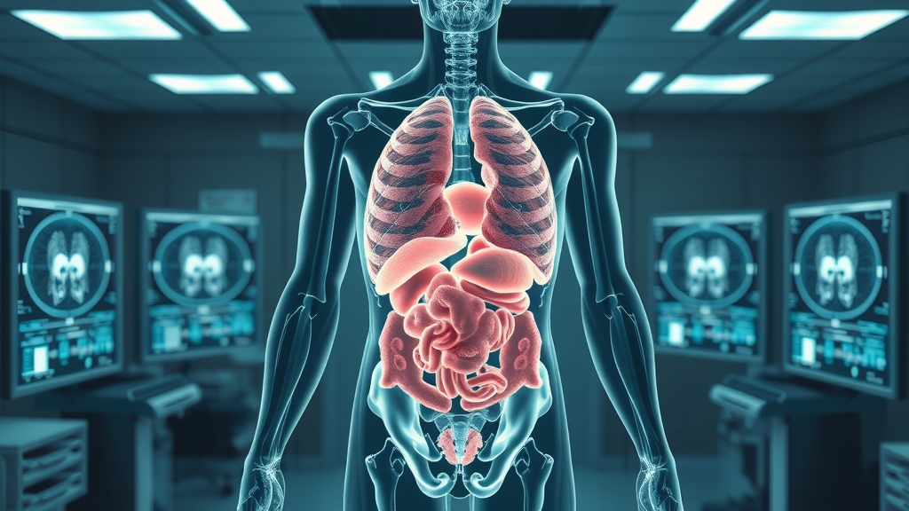
To tackle the pervasive issues in medical image segmentation AI, it’s crucial to first establish a solid foundation. At its core, medical image segmentation involves dividing a medical image (such as a CT scan or MRI) into distinct regions corresponding to different anatomical structures or pathological areas. This process is vital for a wide array of applications, from identifying tumors to planning surgeries, and is powered by advances in both artificial intelligence and computer vision.
Success in this space requires an awareness of both traditional and cutting-edge segmentation methods. While deep learning has revolutionized segmentation tasks, traditional techniques such as thresholding, edge detection, and region growing remain relevant—especially when deep learning models hit roadblocks, such as limited or biased training data. Recognizing the strengths and weaknesses of each approach and knowing when to deploy them is a skill set that separates reliable solutions from those plagued with frustration and unreliability.
Defining Medical Image Segmentation and Its Role in Artificial Intelligence
Medical image segmentation refers to the precise division of medical images into regions or segments that represent different tissues, organs, or pathological areas. This task plays a foundational role in artificial intelligence-powered image analysis. For example, segmenting tumors, organs, or blood vessels in CT and MRI scans is crucial for both diagnosis and treatment planning. Effective segmentation enhances the clarity of input images, allowing AI algorithms to focus on meaningful structures—improving automation and reducing manual annotation in clinical settings.
Artificial intelligence—particularly deep learning approaches such as convolutional neural networks (CNNs)—has taken center stage in automating image segmentation tasks. AI models are trained on extensive datasets of annotated medical images, learning to distinguish between normal and abnormal tissue, or to identify subtle changes not easily recognized by human experts. But this reliance on training data and the inherent complexity of biomedical image segmentation means that even minor data inconsistencies can disrupt model performance. This raises the stakes for careful data curation and critical evaluation of segmentation models in practical, real-world settings.
Core Segmentation Methods: From Deep Learning to Traditional Approaches
Segmentation methods in medical imaging range from classic algorithms—like thresholding and region growing—to modern deep learning models. Traditional techniques typically rely on low-level pixel information, which makes them relatively interpretable but potentially less adaptable to complex variations in biomedical images. In contrast, deep learning models (such as U-Net and Mask R-CNN) learn features directly from data and can capture more intricate patterns. However, they are often “black boxes,” making it difficult to explain segmentation results and diagnose failure modes.
Choosing the right segmentation method depends heavily on clinical requirements, the diversity of available training data, and the need for model interpretability. For rare disease segmentation or when datasets are limited, hybrid approaches that combine domain knowledge with machine learning are gaining traction. These methods help balance the flexibility of AI with the reliability of tried-and-true image analysis techniques—critical for ensuring reliable outcomes in the clinical setting.
| Segmentation Model | Type | Strengths | Weaknesses |
|---|---|---|---|
| Thresholding / Region Growing | Traditional | Simple, interpretable, minimal data requirements | Struggles with noise, poor at handling complex structures |
| Active Contour (Snake) | Traditional | Good for smooth boundaries, interactive adjustment | Sensitive to initialization, limited automation |
| U-Net (CNN) | Deep Learning | High accuracy, robust for biomedical image segmentation, scalable | Requires large annotated datasets, less interpretable |
| Mask R-CNN | Deep Learning | Multi-object segmentation, flexible for varied input image types | Computationally intensive, can overfit limited data |
| Hybrid Models (AI+Rules) | Hybrid | Balances AI learning with domain heuristics, improved interpretability | Requires multidisciplinary expertise to implement |
How Computer Vision Powers Medical Image Segmentation Tasks
Computer vision forms the backbone of automated medical image segmentation. Using mathematical and statistical techniques, computer vision enables AI systems to extract meaningful patterns from complex input images—ranging from CT scans to ultrasound images and beyond. At the core, these techniques empower neural network and deep learning models to recognize minute differences in tissue, shape, and texture that may go undetected through manual review.
The contribution of computer vision extends beyond pattern recognition. By facilitating semantic segmentation—where every pixel is classified into a relevant category—it streamlines image analysis, enhances diagnostic workflows, and supports vital clinical decisions. This synergy between AI and computer vision is ushering in new standards for speed, accuracy, and reliability in clinical diagnostic settings, but it also presents unique challenges that require cross-disciplinary expertise and ongoing scrutiny.
Common Frustrations in Medical Image Segmentation AI and Their Root Causes
Many practitioners approach medical image segmentation AI anticipating rapid, transformative benefits. Instead, they're often met with frustrating setbacks that range from wildly inconsistent segmentation results to the persistent failure of deep learning models on real-world datasets. These difficulties reflect deeper, structural issues within the field—such as the absence of high-quality, representative training data, the inadequacy of existing evaluation metrics, and the lack of transparency in segmentation models. Understanding these sources of frustration is the first step toward building robust and reliable solutions for real-world clinical use.
This section dives into the tangible root causes—why segmentation performance often falls short of expectations, why training data is both a necessity and a liability, and how semantic segmentation sometimes amplifies rather than eliminates errors. By addressing these points head-on, you’ll gain a realistic perspective—and the tools—for overcoming the challenges unique to medical image segmentation tasks.
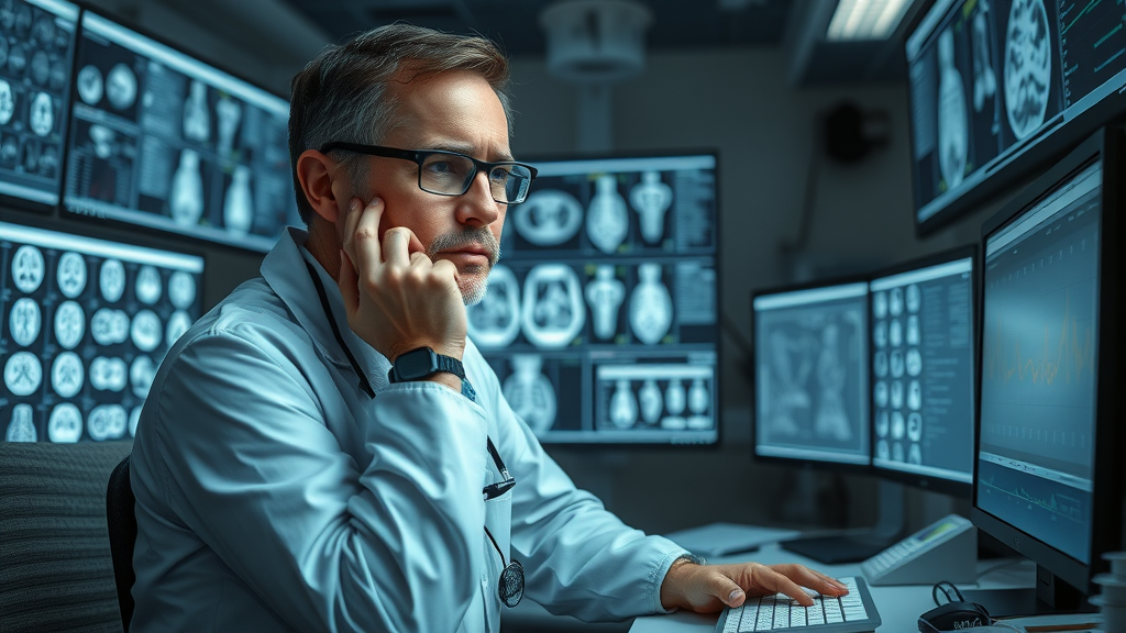
Segmentation Performance: Why Results Frustrate AI Practitioners
Segmentation performance is where the promises and perils of medical image segmentation AI become especially apparent. Practitioners often find that even highly-touted segmentation models underperform on new or unseen datasets. Common causes include a lack of generalizability, overfitting to training data from a single institution, and poor quality control in data labeling. These problems are compounded in sensitive clinical environments, where inconsistent or inaccurate segmentation results can delay or jeopardize patient care.
The fact that no single segmentation model performs optimally across all segmentation tasks highlights a crucial limitation: model evaluation must move beyond artificial benchmarks to reflect real-world complexity. Diverse test datasets, rigorous cross-institutional validation, and ongoing clinician feedback are needed to ensure model performance translates into practical clinical utility. Only by acknowledging these nuances can AI practitioners move beyond surface-level solutions and establish reliable standards for medical image analysis.
The Training Data Dilemma in Medical Images
Training data is the bedrock on which all deep learning and computer vision systems are built; yet, this resource is notoriously difficult to get right in medical imaging. High-quality annotated medical images are expensive and time-consuming to produce. Biases can creep in through overrepresentation of common cases or exclusion of rare diseases and specific patient populations, leading to skewed model performance. Furthermore, privacy regulations and fragmented data sources pose additional barriers to compiling diverse training sets.
When training data does not reflect the full complexity of clinical reality, deep learning models may excel on paper but fail in the real world. The challenge for practitioners is to continually expand, refine, and audit their datasets—incorporating ongoing feedback from both machine learning experts and frontline clinicians. Rigorous attention to dataset construction and curation is as important as algorithm selection for trustworthy image segmentation results.
Semantic Segmentation Pitfalls: Where Deep Learning Fails
Semantic segmentation—where each pixel in a medical image is assigned to a meaningful class—remains a central goal of AI-powered image analysis. However, deep learning models used for semantic segmentation are vulnerable to multiple failure points. These include subtle but critical annotation errors, generalized domain shift problems (when training data differs from deployment scenarios), and a lack of model interpretability, which can mask systematic errors.
These vulnerabilities mean that when segmentation fails in the clinical setting, it can erode trust among medical professionals and jeopardize patient outcomes. To minimize such frustrations, leading practitioners recommend rigorous benchmarking across different segmentation tasks, developing explainable segmentation models, and incorporating human-in-the-loop feedback mechanisms. In the world of medical imaging, transparency, interpretability, and collaboration are not optional—they’re essential safeguards.
Opinion: Why Medical Image Segmentation AI Deserves a Nuanced Approach
"True progress in medical image segmentation will not come from bigger models, but from smarter segmentation methods and honest conversations about limitations."
The race to build “state-of-the-art” models has led many to overlook the significance of workflow design, transparency, and humility in segmentation AI. Oversized neural networks and excessive algorithmic complexity can create an illusion of progress, masking deeper issues related to training data, deployment workflows, and real-world generalizability. What’s needed is a nuanced approach—one that values interpretability, clinician collaboration, and a candid assessment of both successes and limitations.
In this opinion-based analysis, I argue that embedding humility, skepticism, and iterative evaluation into our approach to medical image segmentation AI is not a sign of weakness—it’s the true path to innovation. The best models aren’t just “deep” in architecture; they’re deep in context, interdisciplinary dialogue, and pragmatic deployment. Only then can we move beyond persistent frustrations and transform segmentation models into tools genuinely trusted by clinicians.
Balancing Hype with Realistic Expectations in Medical Image Segmentation
As the hype around AI in healthcare continues to build, it’s easy to assume that ever-larger models will yield ever-better results. However, this mindset can set up both practitioners and decision-makers for disappointment. The reality is: no matter how advanced the artificial intelligence, useful medical image segmentation relies on a combination of sensible workflow design, clinical feedback, and honest performance metrics.
Building trust requires open acknowledgment of AI's limitations and areas for improvement. By inviting skepticism and continuous improvement into the development cycle, we can circumvent the disillusionment that often follows unmet expectations. In my view, this pragmatic optimism—equal parts belief in technology and respect for clinical reality—is the foundation for meaningful innovation in the field.

Why Artificial Intelligence Alone Can’t Solve All Medical Imaging Problems
Artificial intelligence has undeniably advanced medical imaging, delivering breakthroughs in automated diagnosis, segmentation, and workflow integration. Still, expecting AI models to independently resolve all the nuances of medical image analysis is wishful thinking. Deep learning and pattern recognition tools amplify human capability, but they don’t—and shouldn’t—replace the insight and judgment of experienced medical professionals.
The real-world challenges of medical imaging require a symbiosis between human expertise and machine intelligence. Physicians, radiologists, and medical technologists provide critical context, identify edge cases, and catch model errors that elude even sophisticated algorithms. AI models thrive when their limitations are recognized and supplemented by collaborative, iterative processes. In this view, AI is a powerful partner—not a solo problem solver.
Personal Insights: Solving Medical Image Segmentation AI Without Common Headaches
Having encountered and overcome many segmentation roadblocks firsthand, I believe the way forward requires a shift in mindset as much as improved technology. Learning from failures, embracing new segmentation methods, and fostering collaboration lead to more robust and less frustrating AI deployment in clinical contexts. Here are several key insights I’ve found invaluable for sidestepping the enduring headaches in medical image segmentation AI.
The journey to reliable AI-powered image segmentation involves much more than technical horsepower. Genuine progress comes from critically assessing failed models, innovating beyond standard approaches, and creating workflows that prioritize both interpretability and stakeholder buy-in. By integrating clinical feedback and ensuring diversity in training data, you strengthen both the reliability and trustworthiness of your solutions.
Learning from Failed Segmentation Models
Model failure is not just an inconvenience—it's a goldmine of information. Each failed segmentation task, whether due to poor generalization, annotation error, or subtle bias in training data, signals an opportunity for learning and iteration. The best-performing models are products of relentless testing, careful error analysis, and a willingness to rebuild foundational assumptions when needed. It’s essential to move past embarrassment or frustration and view segmentation failure as a road sign guiding you toward improvement.
Emphasizing post-mortem analysis, cross-validation, and interdisciplinary code review helps transform every setback into a stepping stone. In my experience, this strategy is especially effective in healthcare environments, where stakes are high, and every gain in model performance directly translates into improved patient outcomes and clinician confidence.
Innovative Segmentation Methods to Bypass Standard Frustrations
The most innovative segmentation methods often depart from the AI mainstream. Approaches like explainable AI (XAI), hybrid rule-based and machine learning models, and data augmentation using generative networks can alleviate the limitations of black-box models and limited datasets. Prioritizing the interpretability of segmentation results and benchmarking against diverse, real-world medical images can yield more robust and actionable outcomes.
- Prioritize interpretability in segmentation models
- Utilize diverse, representative training data
- Regularly benchmark against multiple segmentation tasks
In contrast to standard deep learning models, these strategies acknowledge that AI is just one piece of the puzzle. When you bring together domain knowledge from clinical experts, data scientists, and informaticians, your segmentation pipeline becomes not only more effective but also more trusted by its end users.
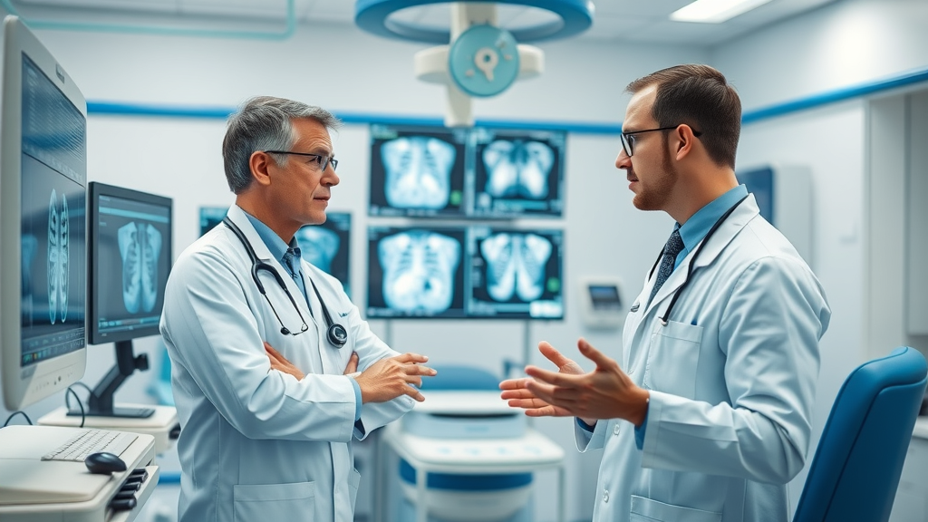
The Importance of Collaboration in Advancing Medical Image Segmentation AI
In the fast-paced world of medical AI, it’s easy to overlook the power of collaboration. Yet, some of the most significant breakthroughs in medical image segmentation occur at the intersection of medical, technological, and human-centered expertise. Diverse teams of radiologists, software engineers, and other specialists contribute broad perspectives, identify blind spots, and drive innovation far beyond what any single discipline can achieve.
Regular interdisciplinary meetings, collaborative data annotation sessions, and open channels for feedback ensure that segmentation methods remain clinically relevant and robust. In my own practice, team-oriented workflows not only accelerate model development but also sharply improve deployment outcomes, minimizing the frustrations that commonly plague siloed AI projects.

Expert Commentary: What Sets Successful Medical Image Segmentation Apart
"Segmentation performance improves not from cutting-edge deep learning tricks alone—it’s the workflow that matters." — Leading Medical AI Researcher
Industry leaders and clinical practitioners consistently point out that the “secret ingredient” to successful medical image segmentation AI is not solely the sophistication of neural networks or the volume of training data. Instead, it’s the thoughtful integration of workflow, human expertise, and pragmatic model validation. Segmentation methods that invite regular clinical input and integrate seamlessly into real-world clinical environments reliably outperform those designed in isolation.
Ultimately, the best segmentation systems respect the context and complexity of healthcare—adapting to new imaging modalities, patient populations, and diagnostic needs. Consistent collaboration and ongoing feedback mean that the technology continues to evolve, minimizing failure points and reducing persistent frustrations.
How Medical Images and Human Expertise Intertwine in Segmentation Tasks
Modern medical image segmentation is a blend of state-of-the-art algorithms and expert interpretation. While AI can parse millions of images and detect subtle patterns, it’s human expertise that ensures clinical relevance and practical value. Radiologists and clinical technologists play a pivotal role in both the initial annotation of training data and the validation of final segmentation outputs.
This partnership is especially critical for complex segmentation tasks, such as those involving rare diseases or atypical anatomical structures. Human input helps tailor model training for nuanced cases that defy statistical norms, reducing error rates and elevating overall model performance. The result is a workflow where error correction, quality assurance, and continual learning are natural byproducts of team-based development.
Case Study: Real-World Success of Medical Image Segmentation AI
Consider the deployment of segmentation models in a state-of-the-art cancer clinic. Initially, the AI was trained solely on well-annotated public datasets, but out-of-sample performance was underwhelming in the clinical setting. By forming a task force of oncologists, radiologists, and software engineers, the team expanded and diversified their dataset, introduced hybrid model strategies, and implemented weekly cross-validation checkpoints.
Within six months, segmentation accuracy for previously problematic tumor types increased by 15%. More importantly, clinicians reported greater confidence in using the segmentation output for treatment planning. This experience underscores the value of cross-disciplinary collaboration, robust workflow design, and a relentless focus on real-world validation—the elements that set truly successful medical image segmentation AI projects apart.
Lists: Top 5 Frustrations and Solutions in Medical Image Segmentation AI
- Inconsistent Training Data Quality → Develop robust data pipelines
- Overfitting to Sample Datasets → Regularize segmentation models and diversify inputs
- Lack of Segmentation Task Generalizability → Test on varied medical image segmentation tasks
- Interpretability Gaps → Employ explainable artificial intelligence approaches
- Workflow Integration Issues → Design solutions with end-user feedback
Leveraging Deep Learning in Medical Image Segmentation AI: Optimism vs. Reality
The arrival of deep learning has radically enhanced the potential for automated medical image segmentation. U-Net, Mask R-CNN, and similar models are now standard bearers for state-of-the-art performance. Yet, their promise is tempered by well-known limitations, including dependency on abundant training data, risk of overfitting, and challenges in model interpretation. In this section, we balance the optimism of deep learning’s transformative power with the reality that it’s not a universal solution for every segmentation task.
Going forward, hybrid approaches—combining deep models with classic segmentation methods and clinician insight—will define the highest-performing, least frustrating solutions in medical AI. The future lies in integrative strategies that acknowledge both the computational strengths of AI and the contextual, interpretative skills of medical professionals.
How Deep Learning Has Reshaped the Segmentation Task
Deep learning’s entry into medical image segmentation has been characterized by explosive gains in accuracy and efficiency. Models like U-Net leverage thousands of annotated input images, learning features and relationships far too complex for traditional algorithms. This has made automation possible even for complicated tasks like multi-organ segmentation and differentiation of overlapping structures. As a result, deep learning has replaced manual annotation as the default for many routine segmentation workflows.
Still, these gains come with caveats. High performance in the laboratory does not automatically translate to consistent results in clinical practice, where data is messy and edge cases abound. Ensuring generalizability, transparency, and adaptability remains a critical concern. This means that while deep learning has replaced human effort in some aspects of image analysis, human oversight is as vital as ever.
Limitations of Deep Learning Approaches for Medical Image Analysis
The Achilles’ heel of deep learning in medical imaging is its reliance on large, high-quality annotated datasets. Many segments of the healthcare industry lack the resources or infrastructure to produce sufficient training data, making these solutions less accessible and potentially less reliable in underrepresented populations. Additionally, black-box model architectures impede understanding of why a given segmentation output was produced, which is problematic in high-stakes clinical environments where explainability is vital.
As a consequence, several initiatives now focus on developing interpretable models, robust post-processing pipelines, and user-friendly annotation tools—efforts designed to make segmentation results transparent and reproducible. Ultimately, the challenge is not just about pushing accuracy metrics higher, but ensuring that segmentation methods fit seamlessly into clinical practice, where they can perform reliably under real-world conditions.
Future Outlook: Integrating Deep Learning with Traditional Segmentation Models
The next generation of medical image segmentation AI will likely be characterized by a sophisticated integration of deep learning with classic image analysis techniques. Hybrid models that leverage domain knowledge—such as anatomical constraints, statistical priors, or clinician-in-the-loop adjustments—are proving to be more resilient, flexible, and interpretable than pure AI approaches.
As segmentation tasks continue to diversify, the synergy between AI and human expertise will set the standard for reliable, low-frustration solutions. Expect future segmentation methods to prioritize interpretability, adaptability, and seamless clinical integration, while retaining the remarkable pattern recognition capabilities of deep neural networks.
Visualizing Medical Image Segmentation AI: Sample Datasets and Approaches
Effective deployment of medical image segmentation AI requires not only high-performing models but also intuitive visualization tools. These help practitioners assess segmentation quality, compare outputs, and identify both successes and problem areas. Here, we explore how side-by-side comparisons and workflow demonstrations can clarify the impact of AI in real-world clinical settings.
Visualizations also play a key educational role, demystifying segmentation processes for both clinicians and patients. Through sample datasets and step-by-step walk-throughs, AI-driven approaches become more accessible, understandable, and actionable.
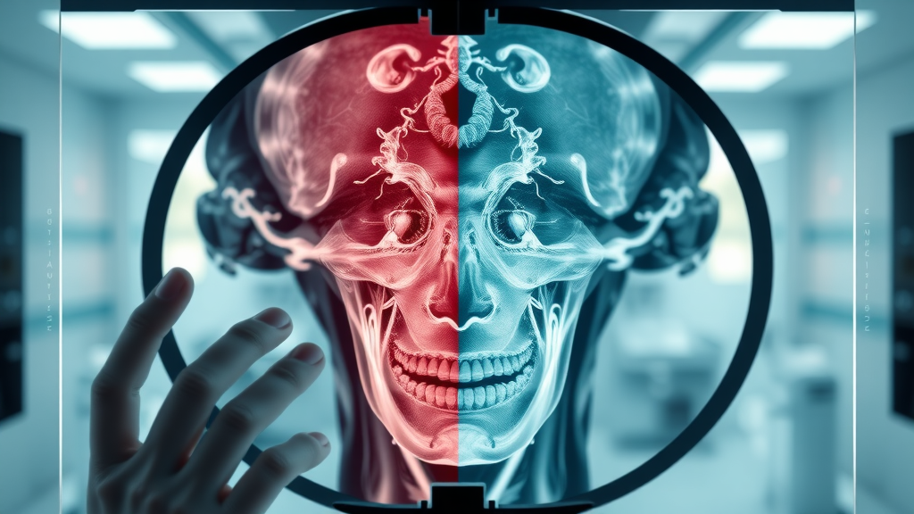
Medical Images Before and After Segmentation: What Experts See
Comparing an original medical scan to its AI-segmented counterpart reveals the power and limitations of medical image segmentation AI. Experts look for clarity of boundaries, correctness of identified regions, and the segmentation model’s ability to generalize across patient populations and modalities. Overlaying color-coded segmentation masks onto input images allows for rapid error identification and informs iterative improvement.
For segmentation methods to be trusted in clinical practice, visual outputs should be both accurate and interpretable. Explaining segmentation results with transparent overlays and stepwise comparisons supports clinician buy-in and enhances patient safety.
Watch a complete walkthrough: From raw input image to final clinical interpretation, this video demonstration details every phase of the AI-driven segmentation process. Expert commentary explains how input medical images are pre-processed, annotated, segmented using deep learning models, and validated by both AI metrics and human experts—offering an inside look at how end-to-end workflows succeed or struggle.
People Also Ask: Medical Image Segmentation AI
Can AI analyze medical images?
Exploring the Capabilities of Artificial Intelligence in Medical Image Analysis
Yes, artificial intelligence—especially deep learning and computer vision—can analyze medical images with remarkable speed and accuracy. AI systems can detect anomalies, segment anatomical structures, and assist clinicians in interpreting complex imaging data. While not a replacement for medical professionals, AI enhances decision-making by rapidly interpreting large volumes of imaging data and highlighting areas of interest. Still, human oversight and validation are essential to ensure reliable diagnostic outcomes.
What is image segmentation in medical imaging?
A Formal Definition and Its Importance in Diagnostic Healthcare
Image segmentation in medical imaging is the process of dividing a medical image into regions representing different anatomical parts or pathology. This enables precise measurement, localization, and diagnosis in applications such as tumor detection, organ delineation, and planning surgeries. Segmentation masks highlight specific tissues or structures, making it easier for clinicians to assess, monitor, and treat patients accurately. As a cornerstone of modern diagnostics, segmentation is foundational for leveraging AI in healthcare.
Is AI going to take over medical imaging?
Realistic Expectations: How AI Complements, Not Replaces, Medical Professionals
No, AI is not expected to take over medical imaging. Instead, artificial intelligence acts as a supplementary tool, streamlining workflows, raising efficiency, and catching patterns that might otherwise escape notice. Clinical judgment, contextual interpretation, and ethical decision-making remain human responsibilities. The most successful deployments harness the strengths of both AI and healthcare professionals—improving outcomes while retaining the irreplaceable value of human expertise.
Which AI technique is commonly used for medical image analysis?
Overview of Deep Learning, Semantic Segmentation, and Other Popular Approaches
Deep learning—specifically convolutional neural networks (CNNs)—is the most commonly used AI technique for medical image analysis. Models like U-Net, Mask R-CNN, and variations of semantic segmentation architectures are widely adopted for tasks ranging from tumor segmentation to organ recognition. Machine learning and pattern recognition techniques also play supporting roles, especially in smaller datasets or when combining image analysis with clinical data.
FAQs: Medical Image Segmentation AI
-
What datasets are commonly used for training segmentation models on medical images?
Popular datasets include the Cancer Imaging Archive (TCIA), NIH Chest X-Rays, LUNA16 for lung nodule analysis, and MICCAI challenge datasets. These provide benchmark cases for evaluating model performance but should be supplemented with institution-specific and diverse data for real-world deployment.
-
How do you evaluate segmentation performance in medical image segmentation tasks?
Metrics like the Dice Similarity Coefficient, Intersection-over-Union (IoU), and Hausdorff Distance are standard for measuring overlap between predicted segmentation masks and ground truth. Clinical validation and real-world testing remain essential to ensuring meaningful performance.
-
How can segmentation methods deal with rare diseases with limited data?
Approaches include data augmentation, transfer learning from related imaging tasks, and the use of explainable or hybrid models. Engaging clinicians in the annotation process and leveraging synthetic data generation are also effective strategies for boosting performance on rare conditions.
Key Takeaways: Medical Image Segmentation AI Opinion Insights
- Medical image segmentation AI is as much about mindset as technology.
- Data quality, segmentation model selection, and workflow design are critical.
- Collaboration and skepticism fuel innovation in artificial intelligence.
Conclusion: Rethinking Medical Image Segmentation AI
"Frustrations in medical image segmentation AI are invitations to innovate, not signs of failure."
The true breakthrough in medical image segmentation AI comes not from chasing the latest algorithmic fad but from honest appraisal, interdisciplinary cooperation, and a relentless focus on practical, real-world results.
Empowering the Next Generation of Medical Imaging and Artificial Intelligence

Watch a roundtable conversation with leading clinicians, AI engineers, and healthcare administrators discussing transformative trends, persistent challenges, and the promise of medical image segmentation AI in the next decade.


Write A Comment