

Over 80% of organizations face bottlenecks in extracting actionable insights due to limitations in automated image interpretation. If you’ve ever struggled with long wait times for results, confusing image analysis outcomes, or feeling like your team isn’t using technology to its fullest, you’re not alone. This comprehensive guide shows you exactly how to overcome common hurdles in automated image interpretation, boost the efficiency of your image analysis pipeline, and implement methods that put you back in control—no matter if you work in healthcare, research, or industry.
"Over 80% of organizations face bottlenecks in extracting actionable insights due to limitations in automated image interpretation."

Opening Insights on Automated Image Interpretation
The explosion of digital images and vast image data across industries has transformed how we process, analyze, and extract meaning. Yet, while the promise of automated image interpretation is enticing—speeding up workflows, reducing manual labor, and providing reproducibility—many organizations find themselves stuck. From the medical image expert trying to detect tumor regions faster, to industrial teams using computer vision for product inspection, bottlenecks persist due to technical, practical, and human challenges. With so many advances in deep learning, neural network tech, and analysis software, taking a critical, informed approach lets you harness the true power of automation without losing oversight or accuracy. This article will demystify automated image analysis from core concepts to practical applications and offer you a roadmap for an efficient, high-quality analysis system.
What You'll Learn About Automated Image Interpretation
- Understanding the foundations of automated image interpretation
- Key comparison points with manual and hybrid image analysis
- Step-by-step methods to refine your image analysis pipeline
- Advanced tools, techniques, and real-world applications
- Best practices for integrating automation in medical, scientific, and industrial domains
Defining Automated Image Interpretation and Its Role in Image Analysis
Automated image interpretation is the use of algorithms and artificial intelligence to mimic and often surpass human analysis when extracting information from images. Unlike traditional manual image analysis, which is labor-intensive and subject to human error, automated systems process a wide range of digital images in a fraction of the time. From feature extraction in cell biology to object detection in computer vision tasks, these methods have redefined efficiency. Technologies like machine learning, neural network architectures, and specialized analysis software enable systems to automatically detect features across diverse data sets, including medical images such as MRI or CT scans, industrial inspection images, and field microscopy data sets for scientific discovery. The flow of data has become more streamlined, empowering experts in health care, manufacturing, and research to achieve reproducible, scalable insights that were previously unattainable.
- Core concepts: automated image, image analysis, image processing
- Types of data and use cases: medical images, computer vision, field microscopy
The Processing Pipeline: How Automated Image Interpretation Works
Understanding the detailed description of the processing pipeline is essential for anyone looking to optimize outcomes in automated image interpretation. The typical workflow begins with image processing—including image denoising and normalization—to ensure high-quality, consistent inputs. Next comes segmentation, where the software delineates specific regions, such as a tumor region in a medical image, or objects on a manufacturing line in computer vision. Feature extraction follows, as algorithms measure and quantify relevant attributes, producing reliable image features from huge volumes of data. Finally, classification systems—powered by deep learning and machine learning methods—assign labels or diagnoses, offering insights that drive clinical workflow, industrial decisions, or scientific discovery. Each stage relies on its own set of algorithms, ranging from general-purpose histogram equalization to sophisticated neural networks tailored for specific use cases, ensuring a seamless flow of data from raw imagery to actionable results.
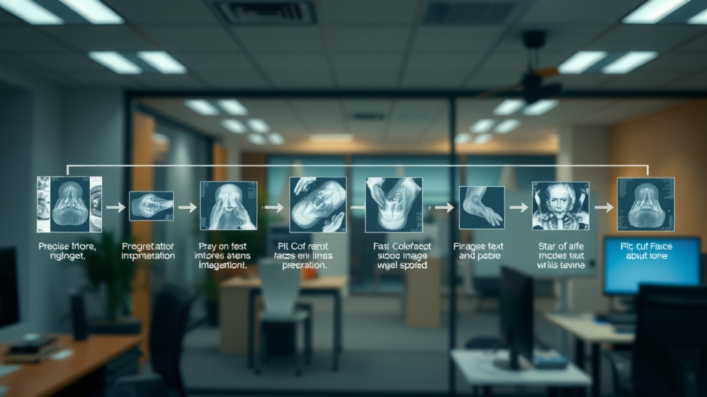
Image Processing and Feature Extraction in Automated Image Analysis
Image processing is the critical first step for all automated image analysis systems. Here, techniques such as denoising (reducing random noise in digital images), normalization, and restoration prepare the raw data for meaningful analysis. Once the images are prepped, segmentation algorithms separate objects or regions of interest—for example, isolating the tumor region in an oncology dataset or individual cells in field microscopy. Feature extraction is where the heavy lifting happens: quantitative statistics such as texture, shape, or intensity patterns are computed automatically, enabling robust distinction between different tissue types, defects, or sample categories.
The automation of feature extraction ensures consistency, reproducibility, and scalability that outpaces manual methods. Instead of slow, subjective measurement, a well-tuned pipeline leverages proven algorithms—like GLCM for texture or U-Net for segmentation—to output a rich set of image features within seconds. Pairing these advancements with a wide range of analysis software, both open-source and enterprise-level, lets teams customize workflows for tasks as diverse as medical image diagnosis, cell lymphoma research, or automated defect detection in industrial quality control.
| Stages of Automated Image Interpretation | Key Tasks | Common Algorithms |
|---|---|---|
| Preprocessing | Denoising, normalization | Gaussian filter, histogram equalization |
| Segmentation | Delineating objects/regions | U-Net, thresholding |
| Feature Extraction | Quantifying data | GLCM, LBP |
| Classification | Assigning labels | CNN, SVM |
Deep Learning and Neural Network Techniques in Automated Image Interpretation
The surge in deep learning and neural network research has revolutionized the landscape of automated image interpretation. Unlike classic rule-based algorithms, neural networks learn directly from data, automatically detecting intricate patterns and features within complex image data. Convolutional neural networks (CNNs) drive state-of-the-art results in a wide range of medical images, from detecting cancer in radiology to highlighting subtle changes in cell structure for biologists. Deep learning accelerates diagnosis, increases consistency, and frequently matches or surpasses human-level performance in image analysis tasks.
These advancements aren’t limited to healthcare. Computer vision applications benefit from neural network models that reliably inspect products, count items, and monitor quality across industrial settings. In field microscopy, deep learning segments and recognizes rare cell types in immense data sets, enabling breakthroughs in diagnostics and life sciences. With a solid processing pipeline, organizations can deploy these technologies to interpret images automatically and improve outcomes, provided that teams also manage data quality, model training, and system oversight to avoid pitfalls such as bias or “black-box” reasoning.

Automated Image Interpretation in Practice: Key Applications
Automated image interpretation is now a cornerstone in many fields, transforming the speed and accuracy of image analysis. Hospitals deploy AI-powered tools to interpret radiology exams, researchers rely on software to scan microscopy slides for cellular abnormalities, and manufacturers use computer vision to spot flaws before products reach the market. The move toward automation has opened doors for more reliable, efficient decision-making that benefits both end users and industry professionals. By leveraging a robust analysis system—from medical imaging to industrial inspection—organizations handle larger case loads and complex image data without sacrificing quality or consistency.
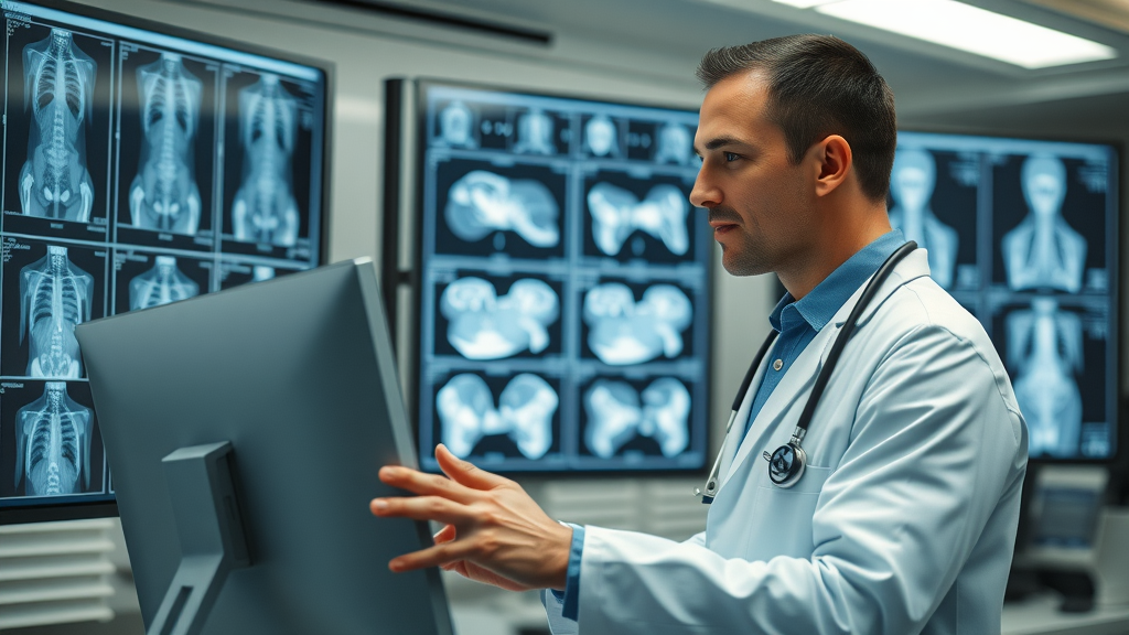
Medical Image Analysis: From Radiology to Oncology
"Automated image interpretation has the potential to detect disease markers faster and more consistently than manual analysis." — Dr. Lin, Radiology Expert
In the healthcare realm, automated image interpretation has dramatically improved the clinical workflow. Systems powered by deep learning and advanced feature extraction can automatically highlight anomalies in MRI, CT, and histopathology images, often surpassing traditional manual review. This is particularly powerful in oncology, where the quick identification of tumor regions can be life-saving. AI models help radiologists identify abnormalities, segment organs, and even predict disease progression, making diagnostic decisions faster, more consistent, and often more accurate. The integration of automated tools in hospital networks in the United States and beyond ensures scalable, reproducible care across a wide range of medical images. Yet, human oversight remains vital—AI predictions must still be validated by medical experts before clinical decisions are made.
Industrial and Scientific Applications: Computer Vision and Field Microscopy
Automated image interpretation extends far beyond medicine. In manufacturing, computer vision systems equipped with cameras and neural network algorithms inspect products, identify defects, verify labels, and monitor machinery health—all in real time. This automation improves accuracy, minimizes waste, and scales rapidly to keep up with high-volume production. Scientific research stands to gain, too: automated field microscopy, for instance, enables quick analysis of vast cell or tissue samples, boosting productivity for biologists and clinical laboratories alike. By replacing subjective manual image annotation with standardized, algorithm-driven processes, a wide range of organizations ensure objective data interpretation and make smarter, evidence-backed decisions every day.

Advantages and Challenges of Automated Image Interpretation
Embracing automated image interpretation comes with clear benefits: workflows become dramatically faster, larger data sets are analyzed with little manual input, and factual, reproducible decisions replace human subjectivity. Whether you’re building a new image analysis system or enhancing a clinical workflow, automation offers scalability, consistency, and often significant cost-effectiveness. However, the journey isn’t without hurdles. Data quality remains a central concern—algorithms trained on limited or biased data can produce misleading or unsafe results. The complexity of deep learning models sometimes leads to “black-box” reasoning, making outcomes difficult to interpret and explain. Maintaining expert oversight and validation steps alongside automation helps strike the vital balance between speed, safety, and accuracy. Ultimately, integrating human experts into the analysis loop ensures the highest level of trust and clinical readiness in both medical and industrial applications.
- Improved speed and scalability
- Potential for objectivity and consistency
- Challenges: Data quality, bias, and interpretability
- Balancing automation with expert oversight
| Benefits | Challenges |
|---|---|
| Efficiency | Data sensitivity |
| Reproducibility | Model bias |
| Cost-effectiveness | Black-box reasoning |

Beyond Automation: Achieving High-Quality Image Analysis Results
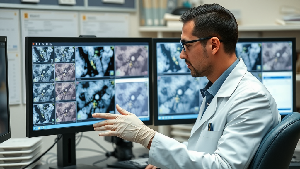
Quality Control in Automated Image Interpretation
Regardless of how advanced your analysis software or automation workflow is, maintaining quality control is essential. Benchmarking automated systems against manual workflows helps identify gaps, outliers, or biases in results. For mission-critical fields like medical image analysis—think detecting cell lymphoma or classifying rare diseases—validation with annotated data sets ensures accuracy and reliability. In research and industry, integrating “human-in-the-loop” systems, where experienced analysts review and validate automated outputs, protects against algorithmic mistakes. This practice leverages the strengths of both automation and expert oversight, helping organizations achieve superior, validated results no matter the data set or use case.
- Benchmarking automated vs. manual workflows
- Validation with annotated datasets
- Integrating human-in-the-loop systems
Improving Your Automated Image Analysis Pipeline
Optimizing your automated image analysis pipeline isn’t a one-time job; it requires ongoing evaluation, collaboration, and innovation. Start by regularly updating training data with new, representative samples to cover a wide range of real-world variability. Continuously benchmark algorithms using both external (public) and internal data to detect drift or degradation in performance over time. Encourage cross-disciplinary collaboration—bringing together data scientists, clinicians, and domain experts—to fine-tune models and ensure output remains relevant for actual decision-making. By nurturing a culture of continuous improvement, you move beyond basic automation to become a leader in extracting maximum value from every digital image your organization encounters.

Key Tools and Software for Automated Image Interpretation
The landscape of automated image interpretation software is growing rapidly, with robust platforms available for every expertise and budget. Open-source solutions such as ImageJ and CellProfiler provide flexible workflows, extensive plugin libraries, and active community support for both biomedical and general-purpose image analysis. Enterprise platforms like MATLAB and Amira offer advanced analytics, seamless integration with large data sets, and support for scripting custom algorithms tailored to unique industrial or scientific needs. Cloud-based options and AI-powered platforms are making high-end automation accessible to organizations of all sizes, while continuously integrating state-of-the-art advances in deep learning and computer vision. For anyone ready to upgrade their analysis system, understanding the strengths, licensing, and feature sets of these tools is crucial for long-term efficiency and success.
- Open-source solutions: ImageJ, CellProfiler
- Enterprise platforms: MATLAB, Amira
- Emerging cloud-based and AI-powered platforms

Watch our introductory video to see how automated image interpretation transforms lab and industrial workflows, featuring real-world cases and easy explanations of the core processing pipeline.
Dive into our in-depth video focusing on how deep learning and neural networks are driving breakthroughs in automated image interpretation for medical image analysis, with tangible examples from current hospitals and research labs.
People Also Ask: Automated Image Interpretation
Is there an AI that can interpret images?
Yes, a wide range of AI systems can interpret images through advanced machine learning and deep learning algorithms. These systems—often referred to as automated image analysis tools—can classify, segment, or detect objects and patterns in medical images, satellite imagery, manufacturing data, and more. Examples include convolutional neural networks (CNNs) for medical diagnostics and vision-based inspection platforms for industry. These AI technologies continue to evolve, increasing accessibility and scalability in image analysis workflows around the world.
What is automated image analysis?
Automated image analysis refers to the process where software interprets digital images without human intervention, usually using artificial intelligence and pattern recognition techniques. This extends from simple measurements (like counting objects) to complex tasks such as diagnosing disease in health care or identifying defects in industrial manufacturing. By leveraging structured processing pipelines—including image processing, segmenting, feature extraction, and automated classification—organizations achieve higher accuracy and efficiency than traditional manual review alone.
Can ChatGPT interpret images?
As of now, ChatGPT itself is primarily designed for text-based tasks and natural language understanding. However, OpenAI and other platforms are advancing multimodal AI models that combine text and image capabilities, allowing for some level of image interpretation when paired with specialized vision components. For comprehensive automated image interpretation, tools specifically designed for image analysis—utilizing deep learning and computer vision algorithms—are more appropriate and widely used in practice.
What are the 7 elements of visual image interpretation?
The seven classic elements of visual image interpretation include: shape, size, pattern, tone or color, texture, shadow, and association. These features guide both manual and automated interpretation by providing quantitative and qualitative clues to identify, segment, and classify objects across a variety of digital images, such as medical diagnostics, satellite imagery, or material science samples.
FAQs: Automated Image Interpretation
-
What are typical data requirements for automated image interpretation?
Robust data requirements usually include well-annotated image data sets that cover all classes of interest, consistent imaging conditions, and high-resolution images where possible. High-quality input ensures algorithms learn the right patterns and generalize well for real-world cases. -
How do you evaluate the accuracy of automated image analysis?
Accuracy is measured by comparing automated results to ground truth annotations using metrics like precision, recall, F1 score, and overall accuracy percentage. Cross-validation on external data and expert review are also crucial for validating the system. -
Which industries benefit most from automated image?
Key benefitting industries include healthcare (radiology, pathology, cell lymphoma detection), manufacturing (quality control via computer vision), life sciences (field microscopy, cell counting), geospatial analysis, and security applications. -
Can human experts override AI interpretations?
Yes, especially in critical applications like medical diagnostics or industrial safety inspections. Many analysis systems include a “human-in-the-loop” design where experts review, validate, or override AI-derived results for maximum accuracy and trust. -
Is automated image interpretation safe for clinical use?
When validated on diverse, well-annotated datasets and supervised by experts, automated image interpretation tools are safe for clinical decision support. Regulatory agencies often require extensive testing and ongoing validation before adoption in clinical workflow.
Key Takeaways: Mastering Automated Image Interpretation
- Automated image interpretation offers transformative efficiency and scalability
- Success depends on understanding algorithms, validation, and application context
- Combining automation with expert insight yields the best analytical outcomes
Conclusion: Take Charge of Your Automated Image Interpretation
By understanding the technology, challenges, and best practices, you can confidently implement automated image interpretation to achieve fast, high-quality results without losing expert control over your workflow.


Write A Comment