Add Row
Add Element
Parallel Health World News Logo
update

Parallel Health World

cropper
update
Add Element
  • Home
  • Categories
    • EcoHealth Trends
    • Healing Naturally
    • Age-Defying Diets
    • Supplement Savvy
    • Mind-Body Synergy
    • Finance and Health
    • Biolimitless
    • Tech Hacks
    • Health & Medicine
    • Political
    • BioBuzz
    • Holistic Rehabilitation Techniques
    • Practitioner Insights
    • AI In Healthcare
  • Featured Business Profiles
September 02.2025
1 Minute Read

Unlock the Magic of ai medical imaging—You’ll Never Go Back

Did you know that nearly 30% of radiologists believe AI medical imaging will redefine their role within five years? That’s not just a statistic—it’s a seismic shift unfolding in hospitals and clinics worldwide. The integration of AI medical imaging isn’t just an upgrade; it’s a revolution reshaping how we detect disease, empower physicians, and improve patient care. Dive into the world where algorithms act as digital diagnosticians, and discover why—once you experience the magic of AI medical imaging—you’ll never want to turn back.

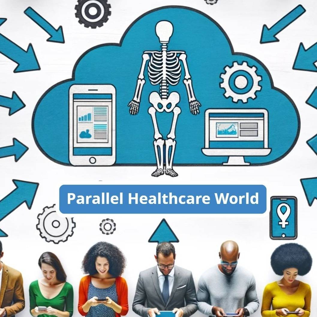
Opening the Conversation: AI Medical Imaging’s Unconventional Rise

AI medical imaging has emerged as a disruptive force in the medical field, rapidly becoming indispensable for diagnosing a wide range of conditions. Traditional radiology relied heavily on a radiologist’s keen eye to interpret X-rays, CT scans, and MRIs, but with the introduction of artificial intelligence, the approach has shifted. Advanced AI algorithms now analyze images at speeds and with precision levels that were once considered science fiction. As these intelligent systems make their way into clinics, they challenge long-held workflows and prompt doctors to reimagine their evolving roles in patient care.

AI in medical imaging offers more than just a technological boost—it paves the way for earlier detection, more accurate diagnoses, and tailored treatment planning. Machine learning and deep learning models, including convolutional neural networks, have demonstrated remarkable progress, guiding the shift from reactive to proactive medicine. The spotlight is now on not just what we can detect, but how much sooner we can intervene, resulting in improved patient outcomes and reducing costly errors. The result is a partnership between human expertise and artificial intelligence that is fundamentally redefining what’s possible in healthcare.

"Did you know that nearly 30% of radiologists believe AI medical imaging will redefine their role within five years?"

The Landscape of Medical Imaging Before Artificial Intelligence

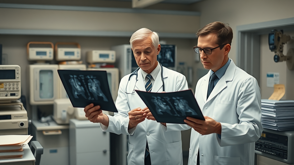
To truly appreciate the transformative power of ai medical imaging, it's important to look back at the era before artificial intelligence entered the medical field. Radiology departments operated under analog systems—think of doctors in white coats, carefully analyzing X-ray films and collaborating over stacks of medical images, each diagnosis relying on years of honed expertise. With limited computational tools, radiologists often faced challenges such as subtle signs being missed or ambiguous shadows leading to inconclusive results. The manual evaluation of medical images was both time-consuming and prone to human error, with early signs of diseases like lung cancer or breast cancer sometimes slipping through unnoticed. Patient care depended heavily on the vigilance and experience of the clinician, but even the best-trained eyes had their limits.

This landscape fostered both innovation and frustration. Without assistive technologies, practitioners juggled swollen workloads, with turnaround times for diagnostic imaging stretching hours or days. As more advanced imaging modalities were developed—CT scans, MRIs, ultrasounds—the sheer volume of medical images skyrocketed. Yet, despite advances in hardware and imaging resolution, interpretation remained a bottleneck. Patient outcomes often hinged on how quickly and accurately radiologists could distinguish benign findings from life-threatening conditions. It became clear that the medical field needed a leap forward to keep pace with the complexity and volume of modern healthcare.

Historical hospital radiology department, radiologists examining X-ray films with analog radiology equipment. AI medical imaging historical context.

Why AI Medical Imaging Matters: A Personal Perspective

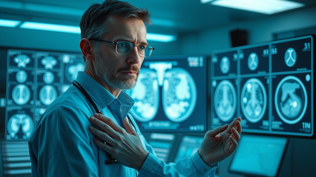
From my vantage point, the real magic of ai medical imaging isn’t just in statistical improvements or faster workflows; it’s in the stories of lives changed and diagnoses made in the nick of time. Having witnessed firsthand how a well-trained AI model flagged a subtle abnormality on a CT scan—a finding that would have taken hours for even an experienced human eye—I am convinced we’re entering a golden age of healthcare innovation. It’s no exaggeration to say that artificial intelligence in medical imaging is saving lives, bridging gaps in care, and alleviating the relentless pressure on physicians.

I've listened to countless practitioners reveal how AI systems have become allies, not adversaries, in their daily routines. The initial skepticism—fueled by fears of being replaced—has given way to cautious optimism as clinicians witness AI’s consistent performance, especially in detecting early signs of diseases like breast cancer or diabetic retinopathy. The greatest benefit is not in replacing expertise, but in augmenting it: physicians can focus on complex cases, patient communication, and decision-making, while AI rapidly processes thousands of medical images for routine findings. The result? Improved patient care, greater workflow efficiency, and a newfound confidence that no subtle anomaly will be overlooked.

As AI continues to integrate into clinical workflows, its impact on both diagnostic accuracy and radiologist workloads is becoming increasingly evident. For a closer look at how artificial intelligence is transforming day-to-day radiology practice and helping ease the burden on healthcare professionals, explore AI’s role in medical imaging and its effect on diagnosis and workloads.

What You’ll Learn From This Exploration of AI Medical Imaging

  • How AI medical imaging enhances patient outcomes and diagnostic accuracy
  • Key breakthroughs in machine learning and deep learning for medical image analysis
  • Controversies and future directions for AI in medical imaging

The Magic Behind AI Medical Imaging: Artificial Intelligence Transforms Medical Image Analysis

Cutting-edge AI interface analyzing a digital medical scan, radiologist observing results. AI medical imaging technology in action.

How Artificial Intelligence Understands and Decodes Medical Images

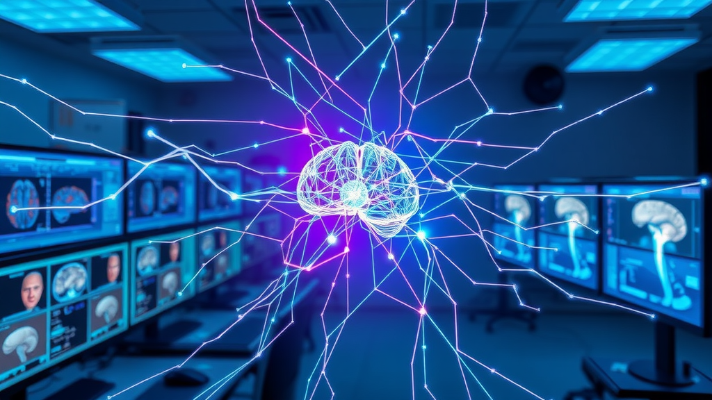
The real leap with ai medical imaging is how artificial intelligence “sees” and understands data. Medical images—once static films or basic digital scans—are now the raw input for deep neural networks and sophisticated machine learning algorithms. These AI systems are trained using vast datasets of annotated images, learning to detect patterns, segment anatomical structures, and spot signs of disease that might evade even the most experienced human observer. Deep learning, powered by convolutional neural networks, excels at complex image classification, distinguishing between healthy tissue and early signs of cancer, stroke, or degenerative diseases. The key is the neural network’s ability to learn from millions of examples, building intuition through repetition, not fatigue.

AI models do more than just point out “abnormal” versus “normal.” They provide heatmaps and probability scores for regions of interest, flagging uncertain findings for further review. In practical applications, this means faster triage for urgent cases and personalized insights for treatment planning. The growing power of AI isn’t a fluke—it’s the product of iterative improvements, relentless innovation, and the constant refining of ai algorithms by interdisciplinary teams of doctors, data scientists, and engineers. Whether analyzing chest X-rays for pneumonia or brain MRIs for subtle tumors, AI is transforming the entire diagnostic journey from grayscale pixels to actionable clinical decisions.

Breakthroughs in Deep Learning and Machine Learning for Medical Imaging

The last decade has seen a surge in breakthroughs at the intersection of deep learning and medical imaging. Advanced algorithms now rival expert radiologists in accuracy, often catching early-stage diseases that once went undetected. For example, convolutional neural networks can sift through massive archives, learning the intricacies of musculoskeletal injuries, identifying microcalcifications in mammograms, or flagging early signs of lung cancer. In many cases, machine learning models outperform traditional image analysis, especially in challenging cases where subtle differences matter most. Notably, AI-enhanced systems have dramatically improved sensitivity and specificity for detecting diabetic retinopathy, enabling earlier interventions and preserving vision for at-risk patients worldwide.

These breakthroughs extend beyond diagnosis—they’re now shaping how medical professionals monitor disease progression, plan surgeries, and predict patient outcomes. From real-time, edge AI-enabled analysis in remote clinics (where expert radiologists may not be available) to cloud-based AI systems that continually plug into global data repositories, the possibilities are only expanding. While the hype is justified by impressive results in controlled studies, the real test of AI medical imaging will be in ongoing, everyday clinical use. Here, the feedback loop between doctors and AI, guided by continuous model improvement, makes each subsequent diagnosis smarter and more reliable.

Personal Stories: Witnessing the Revolution of AI in Medical Imaging

"After seeing AI detect early-stage lung cancer in seconds, I saw the future of patient care."

From Patient Outcomes to Practitioner Confidence: Real-World Impacts

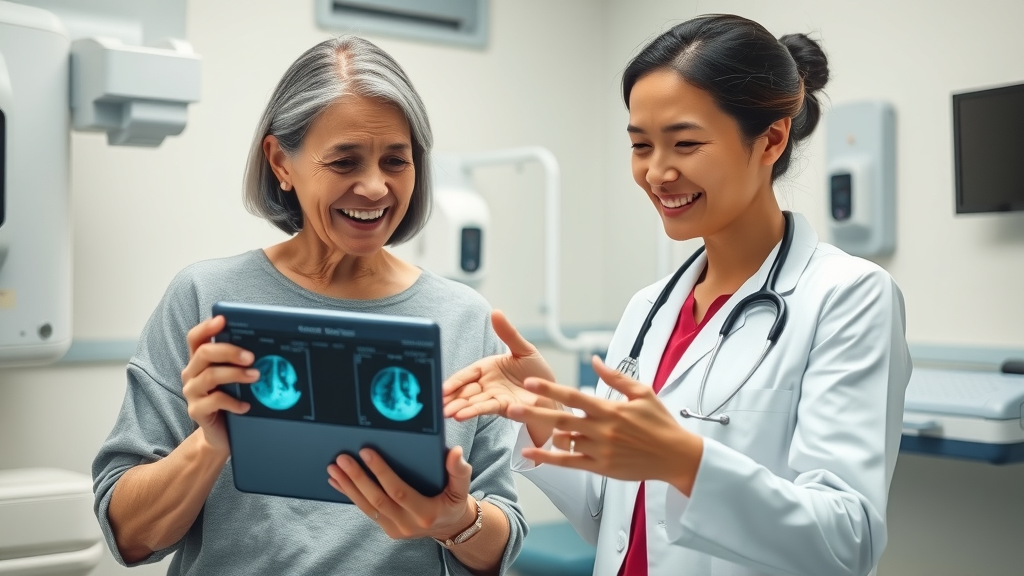
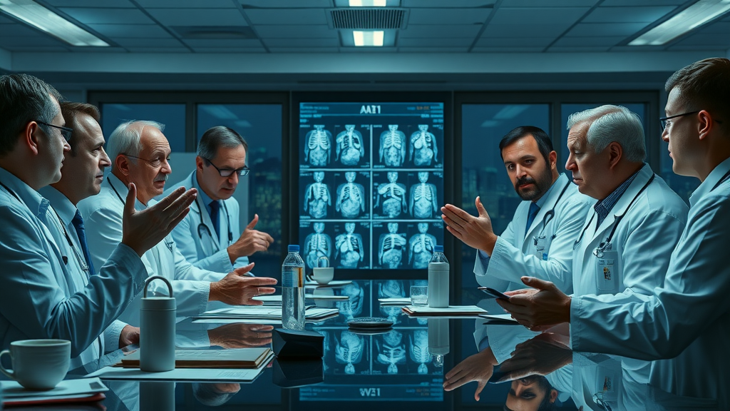
It’s one thing to discuss technology in the abstract; it’s another to witness ai medical imaging at work in a hospital corridor. There’s a quiet but dramatic shift in how care teams operate. In my own experience, I’ve seen patients benefit from earlier interventions for potentially fatal diseases thanks to AI flagging early signs that would have otherwise gone unnoticed. These are not just anecdotes—studies consistently show that ai in medical imaging leads to improved patient outcomes, especially in time-critical cases such as stroke, where every minute matters. The ability of AI models to quickly analyze and interpret medical images minimizes diagnostic delays and allows physicians to initiate life-saving treatments sooner than ever before.

For practitioners, the change is equally profound. Far from feeling threatened, many radiologists now view AI as a colleague—one who never tires, never gets distracted, and is always up-to-date on the latest clinical guidelines. With a second set of AI-enabled “eyes,” doctors report increased confidence in their assessments, and the freedom to focus more on nuanced decisions and patient interaction. While the final call often remains in human hands, the partnership with AI empowers the whole care team, supporting both expertise and empathy in the pursuit of better healthcare.

Watch as a veteran radiologist recounts the moment AI found a critical abnormality in a routine scan, leading to a patient’s lifesaving treatment—and a wholesale change in the doctor’s perspective on the promise of AI medical imaging.

AI in Medical Imaging and Early Detection: A Vital Partnership

How AI Aids Early Detection of Diseases Like Breast Cancer

Early detection saves lives, and nowhere is the impact of ai medical imaging more profound than in screening for conditions such as breast cancer. Using deep learning, AI models can analyze mammograms at scale, highlighting suspicious areas for further examination and vastly improving sensitivity in detecting the disease at its earliest, most treatable stage. Recent advances mean these tools can spot subtle patterns invisible to the human eye, flagging early signs that might otherwise be dismissed as noise. The result ends up being a dramatic reduction in false negatives and improved patient outcomes, especially in populations at elevated risk.

Beyond just detection, these smart systems support radiologists by providing instant, evidence-based second opinions, reducing variability between practitioners, and streamlining reporting workflows. The journey from scan to diagnosis is now shorter, empowering clinicians to start conversations about personalized treatment planning and risk management without delay. As patient care becomes increasingly proactive, AI-driven early detection is poised to become the new standard, particularly for high-volume screening programs where accuracy and efficiency are paramount.

Hopeful patient and radiologist reviewing AI-augmented breast cancer scan on tablet. AI medical imaging for breast cancer early detection.

AI Medical Imaging’s Role in Improving Patient Outcomes

When discussing the promise of ai medical imaging, the most compelling metric is its impact on patient care and patient outcomes. By leveraging advanced algorithms, clinicians gain access to decision support tools that minimize diagnostic errors, expedite treatment initiation, and facilitate ongoing monitoring. In conditions where time is critical—like acute stroke, cardiovascular emergencies, or early-stage cancers—AI’s ability to rapidly process thousands of images and flag subtle changes makes a tangible difference in recovery rates and survival.

Moreover, AI in medical imaging levels the playing field, providing cutting-edge analysis to underserved communities and remote clinics lacking subspecialty expertise. AI algorithms continuously learn from global data, improving with each case and helping close care gaps that demographic or geographical barriers once made insurmountable. With improved accuracy and efficiency, the system reduces unnecessary biopsies and procedures, lessening patient anxiety and overall healthcare costs. In my view, the shift toward AI-augmented diagnostics is one of the most exciting and actionable advances in modern patient care.

Controversial Debates: Is AI Medical Imaging Too Good To Be True?

Doctors and tech experts debating AI results in medical images. AI medical imaging controversies and discussion.

Will Artificial Intelligence Replace Human Radiologists?

No discussion of ai medical imaging is complete without confronting the elephant in the room: will AI systems eventually make human radiologists obsolete? The answer is more nuanced than the headlines suggest. While artificial intelligence and machine learning models have outperformed humans in certain tasks—like pattern recognition and rapid image classification—the gold standard in diagnostic medicine has always required a blend of technical acumen and clinical context. AI can quickly analyze vast troves of medical images, flagging potential concerns, but the final interpretation demands human judgment, empathy, and the ability to integrate complex patient histories.

The professional landscape is shifting from replacement to augmentation. In fact, experts believe that the most effective future lies in AI-human partnerships, where radiologists act as ultimate decision-makers but rely on AI to manage the heavy lifting and identify subtle anomalies. The collaboration helps minimize burnout, speed up diagnoses, and deliver more reliable patient care. Ultimately, as ai in medical imaging becomes embedded in clinics worldwide, clinicians can focus on what they do best: critical thinking, patient communication, and leadership.

The Black Box Problem—Can We Trust AI’s Medical Image Interpretations?

AI medical imaging’s meteoric rise brings a new set of challenges, one of the most profound being the “black box” dilemma. Unlike traditional medical software with explicit criteria and logic, many deep learning systems function as opaque neural networks—making decisions without transparent reasoning. This presents a real concern: how do you trust an algorithm’s diagnosis if you don’t know how it reached its conclusion? In medicine, where lives are on the line, interpretability and accountability are non-negotiable. Regulatory bodies and hospital systems are grappling with the tradeoff: speed versus transparency, automation versus explainability.

Responding to these concerns, researchers are rolling out novel solutions—like generating attention maps that visualize which parts of a medical image influenced AI-driven decisions, and creating traceable audit trails for AI-generated recommendations. Yet, until these systems achieve full explainability, clinicians remain cautious. While many say, “Trusting an algorithm with my diagnosis was unthinkable until the results spoke for themselves,” the push for trustworthy, interpretable AI will only grow as adoption accelerates. It’s a conversation that will define the next decade of AI in medicine.

"Trusting an algorithm with my diagnosis was unthinkable until the results spoke for themselves."

Cutting-Edge Technologies: Machine Learning, Deep Learning, and Medical Imaging

Key Advances in Machine Learning for AI in Medical Image Processing

The heart of recent leaps in ai medical imaging is a set of rapid advancements in machine learning. Unlike past rule-based systems, modern machine learning approaches adapt and improve as they are fed new data. Convolutional neural networks (CNNs), for instance, have been engineered to mimic aspects of human visual perception, providing near-human—or sometimes superhuman—accuracy in image classification tasks. These networks can segment tumors, measure organ volumes, or even quantify subtle biomarker changes across millions of pixels. Another edge AI advancement is the integration of federated learning, enabling the training of robust ai models across multiple hospitals without sharing raw patient data, thus maintaining privacy.

Such advances have made it possible to deploy AI systems across a spectrum of imaging applications: from triaging head trauma in emergency settings to flagging diabetic retinopathy in ophthalmology clinics. The result is a practical toolkit for radiologists and physicians, offering both unprecedented speed and accuracy. The collaboration between advanced ai, patient data security, and continuous model refinement ensures these systems remain relevant and safe. As the field moves forward, the interplay between machine learning, improved algorithms, and diverse datasets will only make AI in medical imaging more powerful and accessible.

Complex neural network visualized as 3D glowing matrix overlaid on medical images. Machine learning and deep learning advances for AI in medical image processing.

How Deep Learning Revolutionizes Complex Medical Imaging Challenges

What sets deep learning apart in the realm of ai medical imaging is its ability to tackle intricate challenges that foiled traditional tools. Deep neural networks don’t just follow pre-written rules—they develop their own methods for parsing and interpreting vast, complex medical datasets. This flexibility is invaluable in tasks such as detecting micro hemorrhages in brain images, isolating subtle pulmonary nodules, and segmenting overlapping anatomical structures. By leveraging large volumes of annotated images, deep learning models identify invisible-to-the-eye cues that can indicate the difference between a benign or malignant lesion, an old injury or a new one.

An especially promising area is the use of generative models, a form of deep learning that can create synthetic medical images for training or testing, expanding limited datasets for rare conditions. These innovations not only enhance diagnostic accuracy but also democratize access to advanced diagnostics, even in areas with few specialists. The versatility and adaptability of deep learning have placed it at the forefront of transformative technology in medical diagnostics, allowing ai in medical imaging to handle the diverse and ever-evolving challenges of patient care.

Lists: Where AI Medical Imaging Shines—and Where It Falters

  • 5 Biggest Success Stories of AI in Diagnostic Medical Image Analysis
    • AI detection of early-stage breast cancer on digital mammograms, resulting in higher survival rates.
    • Automated identification of diabetic retinopathy in retinal scans, preventing vision loss for millions.
    • Rapid triage of brain CT scans for stroke diagnosis, enabling faster intervention and improved recovery.
    • Detection of early lung cancer on low-dose CT, providing timely treatment options for at-risk patients.
    • Streamlining skeletal fracture detection in X-rays, reducing diagnostic errors and patient wait times.
  • 3 Major Limitations Still Facing AI Medical Imaging Today
    • Lack of transparency in deep learning models, contributing to the “black box” problem and regulatory hurdles.
    • Bias in training data leading to inconsistent results across different demographic groups and healthcare settings.
    • Data privacy and cybersecurity concerns, especially with large-scale sharing of patient data for AI model training.

Table: AI Medical Imaging vs. Traditional Methods

AI Medical Imaging Traditional Methods
Diagnostic Accuracy High, especially for early detection (rivaling or exceeding expert radiologists in some applications) High but variable—subject to fatigue and human error, accuracy depends on clinician experience
Speed Rapid, real-time analysis—often minutes or less per case Slower—manual review can take hours or days per case
Cost Potentially lower long-term, improves with scale and automation Can be high due to labor, repeat imaging, and error correction
Patient Outcomes Improved through earlier detection, fewer missed diagnoses, and tailored treatment planning Improved but limited by availability of specialists and variable workload

People Also Ask: Common Questions About AI Medical Imaging

How is AI being used in medical imaging?

Answer: AI is used in medical imaging to automate detection of abnormalities, segment anatomical structures, and support diagnostics through rapid interpretation of X-rays, MRIs, and CT scans, utilizing machine learning and deep learning algorithms.

Can AI generate medical images?

Answer: Yes, AI can generate synthetic medical images for research, training algorithms, and even creating diagnostic imaging scenarios, leveraging advanced generative models in deep learning.

Will AI take over medical imaging jobs?

Answer: While AI medical imaging streamlines workflows and improves accuracy, most experts believe it will augment rather than replace radiologists, enabling better patient care and outcomes.

How accurate is medical imaging AI?

Answer: AI-powered medical imaging has shown accuracy levels rivaling, and sometimes surpassing, experienced radiologists in detecting certain conditions, especially in early detection and screening programs. However, results vary by application and dataset.

Watch a video demonstration showcasing the workflow of AI medical imaging, from image acquisition to AI-assisted diagnosis. See on-screen overlays of AI-generated insights and witness how radiologists interact with digital results in real time.

FAQs About AI Medical Imaging

  • What diseases benefit most from AI medical imaging?
    Diseases that benefit most include breast cancer, lung cancer, diabetic retinopathy, stroke, and musculoskeletal injuries. AI excels in early detection and rapid triage for these and similar conditions.
  • Is patient data privacy at risk when using artificial intelligence in healthcare?
    Like any digital health solution, AI medical imaging must address data privacy risks. Advances in federated learning and strong encryption help mitigate these concerns, but robust security frameworks and regulatory compliance remain essential.
  • What are the regulatory challenges for AI in medical imaging?
    Challenges include the need for transparent algorithms, validated performance across diverse populations, and continuous oversight. Agencies are evolving standards to keep pace with rapid innovation, but the process is ongoing and complex.

Key Takeaways: The Future of AI Medical Imaging

  • AI in medical imaging is accelerating accurate diagnostics and patient care.
  • Both machine learning and deep learning are catalysts for change in medical imaging.
  • Ethical, regulatory, and technological debates must be addressed for mainstream adoption.

Conclusion: Why You’ll Never Go Back After Experiencing AI Medical Imaging

Experience the future of medicine: with ai medical imaging, diagnostics become more accurate, faster, and accessible—making the impossible routine and reshaping the standard of care forever.

If you’re inspired by the rapid evolution of AI in medical imaging and want to deepen your understanding of its broader impact, there’s even more to discover. The journey doesn’t end with improved diagnostics—AI is also transforming how radiology teams collaborate, manage workloads, and deliver care at scale. For a strategic perspective on how artificial intelligence is shaping the future of healthcare and redefining the radiologist’s role, take the next step and read about AI’s expanding role in medical imaging and its influence on the healthcare landscape. Unlock new insights and see how the synergy between technology and human expertise is setting the stage for the next era of patient-centered innovation.

AI In Healthcare

1 Views

0 Comments

Write A Comment

*
*
Related Posts All Posts
09.09.2025

Act Now: The Window to Fix Your AI in medical image processing Is Closing

Did you know that while more than 80% of hospitals plan to deploy AI in medical image processing by 2025, only a third are confident their systems are truly robust or free from bias? This shocking gap isn't just a statistic—it's a loud wake-up call for everyone from radiologists to hospital CIOs. Right now, we are at a crossroads: act swiftly and fix the cracks in AI algorithms and oversight, or risk compromising both patient care and future innovation. In this comprehensive editorial, we’ll explore why the clock is ticking, the challenges that remain, and why taking decisive steps today will shape the next era of medical imaging. A Startling Reality: The Current State of AI in Medical Image Processing The landscape of AI in medical image processing is rapidly transforming, yet lagging behind in critical areas like reliability, transparency, and bias mitigation. While artificial intelligence promises enormous improvements—such as faster diagnostics, optimized treatment planning, and even predictive analytics for diseases like lung cancer or breast cancer—many deployed AI systems still struggle with systemic weaknesses. These include insufficiently diverse imaging data, unexplained neural network decisions, and inconsistencies in regulatory oversight that ultimately place patient outcomes at risk. Today, leading hospitals and clinics are in the process of integrating AI tools for everything from image segmentation to anomaly detection. However, the rate of actual adoption is not keeping pace with the promises of deep learning and machine learning. As a result, many institutions are caught between the accelerating pressure to modernize and the reality that their AI algorithms are still nascent, often opaque, and sometimes inconsistent across different medical images. The urgency here stems from the possibility that, unless addressed now, these limitations could solidify and create long-term barriers to equitable, safe, and effective healthcare. Unveiling the Numbers: AI Adoption in Medical Imaging "Over 80% of hospitals plan to deploy AI in medical image processing by 2025, yet only 30% have robust, bias-free systems ready." These figures underscore a dangerous dichotomy in the medical imaging field. As medical imaging tech advances at an unprecedented rate, the groundwork underpinning successful, fair implementation of AI in medical imaging is being laid unevenly. This discrepancy means many health organizations face the risk of deploying AI solutions that could perpetuate existing biases in imaging data, compromise diagnostic accuracy, and impact patient care—especially for underrepresented groups. Notably, the current momentum among healthcare institutions to implement AI tools stems from the clear benefits AI algorithms and convolutional neural networks promise: scalable diagnostic platforms, more accurate radiological reads, and the ability to handle a deluge of digital medical images. Yet, with so much at stake, the industry must confront the fact that progress in artificial intelligence alone cannot guarantee better patient outcomes without a concurrent commitment to mitigating bias, ensuring data representativeness, and increasing explainability in deep learning systems. What You'll Learn About AI in Medical Image Processing Why urgency matters: the shrinking window for reliable AI in medical image processing Major obstacles and opportunities shaping AI in medical imaging Expert insights and government perspectives on artificial intelligence in healthcare imaging Actionable steps for institutions, radiologists, and decision-makers Why the Rush? The Shrinking Window to Fix AI in Medical Image Processing Decisive action is needed now because the technological evolution in medical imaging is outpacing the careful assessment, standardization, and regulation required to ensure safe deployment of AI systems. As momentum builds—with new learning algorithms and AI tools rolled out at an increasing clip—the window to implement robust, bias-resistant frameworks is narrowing. If stakeholders wait, systemic flaws could become entrenched, eroding both diagnostic accuracy and public trust. The opportunity to make meaningful course corrections is truly time-sensitive. Investment in better imaging data curation, integration of human eye oversight, and improvement of explainability in AI algorithms needs to keep pace with advances in machine learning. Otherwise, hospitals stand to inherit AI systems that are powerful yet fundamentally limited—putting patient outcomes and even regulatory compliance on the line. Technological Momentum: Medical Imaging Outpacing Standards Clinical imaging innovation is accelerating rapidly with widespread use of deep learning, machine learning, and convolutional neural networks for analyzing complex medical images. Algorithms are now capable of identifying early signs of diseases like breast cancer and lung cancer faster than ever, promising a step-change in patient care. However, this technological velocity often surpasses the pace at which ethical, regulatory, and technical standards are updated—another risk factor that demands attention. For instance, while an AI tool might achieve spectacular diagnostic accuracy in a research setting, its performance can drop dramatically in the real world if imaging data used for training is not diverse enough. This is why technology-driven environments need mechanisms for continuous validation and recalibration—without these, the gap between capability and trustworthiness in medical imaging will only widen. Systemic Risks: Bias, Error, and Liability in Algorithmic Medical Image Analysis One of the gravest concerns in deploying AI algorithms for medical image analysis is the risk of ingrained bias—whether in the imaging data used to train neural networks or in the modeling assumptions of the AI system itself. These biases can lead to disparate accuracy rates across demographics, making the role of continuous human supervision and standardized testing indispensable. Errors in AI systems used for medical imaging introduce unique liability and ethical questions that few institutions are fully equipped to handle. Beyond individual misdiagnoses, the propagation of unchecked bias or error means at-scale harm to entire patient populations. To ensure improved patient outcomes, leaders in healthcare must double down on building transparent, auditable, and well-governed AI in medical solutions before mass adoption is complete. How AI in Medical Image Processing Is Reshaping Healthcare The introduction of AI in medical imaging is fundamentally altering the future of diagnostics, patient tracking, and care delivery. Using machine learning and deep learning algorithms, these systems can process vast quantities of medical images rapidly, identifying subtle patterns that the human eye might overlook. From reducing turnaround times for critical image reads to helping personalize treatment planning, AI-driven workflows are making real differences—but only when implemented judiciously and ethically. Particularly, advances in image segmentation, feature extraction, and AI-driven anomaly detection already demonstrate how neural networks and convolutional neural networks can augment radiological interpretation. However, realizing the full promise of AI in medical image processing still hinges on balancing automation with ongoing human oversight and tackling challenges around explainability, generalizability, and equitable training data. Case Study: Deep Learning Advancements in Breast Cancer Detection One illustrative example comes from breast cancer screening, where deep learning models are now capable of identifying malignant features on mammograms with accuracy rivaling—or sometimes exceeding—experienced radiologists. Here, AI algorithms trained on vast banks of medical images can spot early lesions, reduce diagnostic subjectivity, and help prioritize follow-up for suspicious findings. Research has shown this can lead to earlier interventions and, in many cases, improve patient outcomes especially for hard-to-detect cases. Yet, it’s essential to note that these systems often struggle when exposed to image variations outside their training set—for instance, data from different types of scanners, or new population groups. To maximize real-world benefits of AI in medical imaging, models must be continually updated, validated, and overseen by clinical experts to avoid missing rare pathologies or amplifying existing disparities in diagnostic accuracy. Machine Learning & Imaging Data: Revolutionizing Patient Outcomes Machine learning runs on the backbone of well-labeled, representative imaging data. When properly harnessed in medical image processing, these algorithms excel at recognizing subtle, complex features invisible to even experienced radiologists. For instance, they detect nuances in lung cancer nodules or microcalcifications in mammography scans, facilitating early signs detection and better treatment planning. The use of learning algorithms—especially convolutional neural networks—has improved performance in automated image segmentation, organ delineation, and quantification of tumors, directly leading to improved patient outcomes. But this progress also relies on the quality, diversity, and scope of input image data, and highlights the critical need for ongoing data curation and model retraining as clinical scenarios evolve. Artificial Intelligence and Human Oversight: The Delicate Balance in Medical Imaging While AI brings computational power and pattern recognition capabilities beyond human reach, their integration into medical image interpretation is never a case for sidelining clinicians. Instead, the next generation of AI in medical image processing is defined by thoughtful collaboration between AI systems and human radiologists, leveraging the strengths of both while mitigating the risk of relying solely on automated outputs. This human-AI partnership is critical for reducing errors. Human experts catch context-specific subtleties and provide real-time feedback on algorithmic performance, while AI automates the detection of well-characterized patterns, quantifies subtle features, and quickly processes massive image sets. This synergistic approach is central to scalable, high-quality patient care in a rapidly digitizing healthcare environment. Medical Image Interpretation: What Machines Miss and Humans Catch Even the most sophisticated artificial intelligence models can stumble on atypical presentations or rare pathologies that aren't well-represented in their training imaging data. Radiologists contribute essential contextual and experiential knowledge, identifying clues that an AI system might miss, such as subtle background abnormalities or non-standard imaging artifacts. The result is a really robust safety net—one that leverages the precision and speed of AI algorithms with the nuanced judgement of the human eye. Ultimately, the most effective solutions aren’t about replacing radiologists, but augmenting them. This hybrid approach is especially essential for complex diagnoses, uncertain cases, and evolving disease presentations where the context, history, and whole-patient perspective matter as much, if not more, than pure image analysis. Patient Care Considerations: From Image Analysis to Improved Patient Outcomes Patient care extends beyond accurate image reads. Integration of AI in medical image processing impacts everything from faster triage and streamlined treatment planning, to reducing unnecessary procedures and ensuring equitable access to leading-edge diagnostics. AI-driven workflows can shorten waiting times, optimally route patients to the right experts, and even provide second-read support—all which directly impact patient outcomes. But this newfound efficiency must never overshadow the human touch essential to medicine. Empathy, clear communication, and holistic understanding should remain at the center, guiding both the development and deployment of AI tool solutions. Only by prioritizing patient care at every step can AI fulfill its promise as a genuine improvement in healthcare—not just for the technology’s sake, but for people’s lives. State of the Market: AI Tools in Medical Imaging Today The market for AI in medical image processing is now home to a growing array of AI tools that claim to automate everything from simple measurements to complex lesion detection. Global investment and VC interest reflect the sector’s transformative potential, but this proliferation also brings a sea of options and little standardization—making selection, integration, and validation difficult for healthcare leaders. Vendors tout solutions for specific specialties—like AI-driven breast cancer detection, lung cancer screening, or organ segmentation—but not all tools are created equal. Differences in training data scope, regulatory approval (such as FDA clearance), and performance transparency challenge hospitals to separate robust clinical partners from experimental offerings. As the market matures, user-friendly interfaces, integration with existing PACS/EHR, and real-world validation data are quickly emerging as essential markers of reliable AI for medical imaging. Market Leaders: The AI Tool Landscape Several companies stand out within the AI tool market, each targeting different modalities and specialties. Leaders offer end-to-end AI platforms capable of handling a variety of medical images—CT, MRI, ultrasound, and digital x-rays—while ensuring interoperability and security of patient data. These solutions are shaped by their ability to demonstrate clear improve patient outcomes, gain regulatory clearance, and offer support for continuous improvement as imaging protocols evolve. Other challengers take a more focused approach, creating best-in-class solutions for single applications such as image segmentation of brain tumors or early detection in breast cancer screenings. Evaluating these tools requires rigorous side-by-side testing for diagnostic accuracy, usability, integration ease, and transparency of the underlying AI algorithm. Successful deployment depends as much on organizational readiness to adopt and monitor these AI tools as on the technology itself. Barriers to Broad Adoption in Medical Image Processing Despite the range of available tools, comprehensive adoption of AI in medical imaging faces persistent obstacles. Core challenges include inconsistent standards for imaging data, a lack of universally accepted protocols for training deep learning systems, and ongoing concerns about how “black box” AI algorithms reach their decisions. Patient privacy and data-sharing constraints complicate the assembly of diverse, high-quality datasets necessary for robust model development and validation. Additionally, many clinical deployment hurdles remain—from integration with existing radiology workflows to ensuring AI system outputs are interpretable and actionable by human experts. Meeting these challenges will require concerted collaboration between industry, regulators, and medical professionals—and action must be taken now before today’s limitations become tomorrow’s unfixable defects. Comparison of Leading AI in Medical Image Processing Tools AI Tool Specialty/Use-Case Strengths Weaknesses Regulatory Status Al Detect Pro Breast Cancer Screening High sensitivity, fast workflow integration Black box decisions, limited cross-population data FDA cleared PulmoNet Lung Cancer Nodule Detection Advanced deep learning, multi-modal support Requires large training datasets, explainability issues Pending approval CardioScan AI Cardiac MRI/CT Analysis Detailed segmentation, clinician dashboard Integration challenges, slow on legacy hardware EU MDR/CE certified NeuroVision Brain Tumor Localization State-of-art neural networks, intuitive UI Lack of pediatric dataset diversity FDA submitted Expert Perspectives on AI in Medical Image Processing "Human-AI collaboration is the only scalable solution to current bottlenecks in patient care and medical imaging." — Dr. Elaine Park, Radiologist Expert consensus across radiology, data science, and health informatics highlights the non-negotiable need for collaboration. Leading physicians stress that AI tool outputs must always be interpreted within clinical context, with transparent feedback loops so AI algorithms can be improved and revalidated in real time. Meanwhile, data scientists advocate for more representative and diverse imaging data, and hospital administrators urge for clearer regulatory pathways to allow safe but agile innovation. Government & Regulatory Viewpoints on Artificial Intelligence in Medical Imaging Government agencies and regulators globally are grappling with how to foster safe innovation in AI in medical imaging. The FDA, EMA, and other health bodies are working to define clear pathways for evaluating deep learning models and approving new AI tools for clinical use. A major challenge is keeping regulations responsive to the pace of technological change without compromising on core tenets: safety, equity, and patient data privacy. Increasingly, policy frameworks emphasize transparency, demands for post-market surveillance, and calls for algorithmic explainability—requiring clear documentation on how AI system decisions are reached. These standards aim to protect patient welfare and public trust, while enabling responsible and ethical scale-up of artificial intelligence in medical imaging. Key Challenges Facing AI in Medical Image Processing Data bias in imaging data: Non-representative datasets can result in AI algorithms that underperform for certain populations. Lack of standardized deep learning protocols: Inconsistent model training impacts reliability and comparability. Black box algorithms and explainability issues: Clinicians and patients need to understand how AI systems reach medical decisions. Patient data privacy and ethical considerations: Innovative AI tool development must always uphold the sanctity of patient confidentiality. Patient Outcomes & The Real-World Impact of Imperfect AI Inequities and imperfections in AI in medical image processing can have far-reaching consequences on patient care and trust. When AI algorithms misinterpret images due to poor data quality or systemic bias, patients can be subject to misdiagnosis, delayed treatment, or unnecessary procedures—especially in high-stakes contexts like breast cancer screening or lung cancer evaluation. The potential for improved patient outcomes is immense, but only if all players—technologists, clinicians, and policymakers—move quickly to address known flaws. Redoubled efforts to ensure transparency, accuracy, and ethical development will enable AI in medical imaging to fulfill its promise as a force for good, rather than a source of new risk. An animated explainer showing how deep learning algorithms analyze medical images, highlighting collaboration between AI and radiologists. Explore the dual nature of AI in medical image processing—unrivaled opportunity and pressing risk—in this essential perspective video. People Also Ask: How Is AI Being Used in Medical Imaging? AI in medical image processing is revolutionizing diagnostics by enabling faster, more accurate interpretation of radiology scans, segmentation of tumors, and pattern recognition in complex imaging data. By integrating deep learning and machine learning, AI tools help radiologists improve patient outcomes and reduce diagnostic errors. People Also Ask: Can AI Generate Medical Images? Yes, AI can generate synthetic medical images for training, research, and managing data scarcity. Generative models and deep learning allow artificial intelligence to create realistic medical image datasets for safer, more robust algorithm development. People Also Ask: What Is the Role of AI in Healthcare Image? The role of AI in healthcare imaging spans early disease detection, workflow automation, patient triage, and enhanced image analysis—all of which contribute to better patient care and resource allocation in clinical settings. People Also Ask: Can AI Do Image Processing? AI excels at image processing, particularly in medical imaging, where machine learning algorithms automate segmentation, noise reduction, and feature extraction, facilitating more accurate diagnoses and treatment decisions. Essential Steps Forward: What Needs Fixing in AI for Medical Image Processing Instituting data standardization and reduction of bias Implementing ongoing human oversight Improving regulatory frameworks for artificial intelligence Prioritizing patient outcomes over performance metrics FAQs About AI in Medical Image Processing What are the main limitations of AI in medical image processing? Primary limitations include data bias, lack of training data diversity, insufficient explainability of how AI algorithms reach conclusions, and challenges with integration into existing clinical workflows. Overcoming these requires collaboration, rigorous validation, and ongoing oversight. How is deep learning different from traditional machine learning in medical imaging? Deep learning leverages layered neural networks that automatically extract complex features from imaging data, enabling more nuanced pattern recognition compared to traditional machine learning, which often requires manual feature selection. This allows deep learning to solve harder medical imaging challenges but also demands much larger datasets. Are AI tools FDA approved for clinical use in medical imaging? Some AI tools for medical image processing are FDA approved, particularly those with robust clinical validation and safety data. However, many are still under review or in limited use based on specific regulatory pathways. Always check the status and clinical evidence before clinical deployment. How does AI improve patient care in radiology? AI in medical imaging boosts patient care by enabling faster, more consistent image reads, early detection of disease, reduction of human error, optimized treatment planning, and helps ensure better allocation of clinical resources. Most importantly, it supports clinicians in making more informed and timely decisions. Key Takeaways: The Urgency for Robust AI in Medical Image Processing The growth in AI in medical image processing offers immense potential but also introduces urgent challenges. Stakeholders must act now to ensure safe, equitable, and effective implementation. Collaborative regulation, transparency, and patient-centered goals are non-negotiable. Conclusion: Don’t Let the Window Close on AI in Medical Image Processing Ignoring the urgency could undermine both patient care and technology’s promise—stakeholders must act decisively today.

09.08.2025

Tired of automated image interpretation Holding You Back? Here’s How to Take Control

Over 80% of organizations face bottlenecks in extracting actionable insights due to limitations in automated image interpretation. If you’ve ever struggled with long wait times for results, confusing image analysis outcomes, or feeling like your team isn’t using technology to its fullest, you’re not alone. This comprehensive guide shows you exactly how to overcome common hurdles in automated image interpretation, boost the efficiency of your image analysis pipeline, and implement methods that put you back in control—no matter if you work in healthcare, research, or industry. "Over 80% of organizations face bottlenecks in extracting actionable insights due to limitations in automated image interpretation." Opening Insights on Automated Image Interpretation The explosion of digital images and vast image data across industries has transformed how we process, analyze, and extract meaning. Yet, while the promise of automated image interpretation is enticing—speeding up workflows, reducing manual labor, and providing reproducibility—many organizations find themselves stuck. From the medical image expert trying to detect tumor regions faster, to industrial teams using computer vision for product inspection, bottlenecks persist due to technical, practical, and human challenges. With so many advances in deep learning, neural network tech, and analysis software, taking a critical, informed approach lets you harness the true power of automation without losing oversight or accuracy. This article will demystify automated image analysis from core concepts to practical applications and offer you a roadmap for an efficient, high-quality analysis system. What You'll Learn About Automated Image Interpretation Understanding the foundations of automated image interpretation Key comparison points with manual and hybrid image analysis Step-by-step methods to refine your image analysis pipeline Advanced tools, techniques, and real-world applications Best practices for integrating automation in medical, scientific, and industrial domains Defining Automated Image Interpretation and Its Role in Image Analysis Automated image interpretation is the use of algorithms and artificial intelligence to mimic and often surpass human analysis when extracting information from images. Unlike traditional manual image analysis, which is labor-intensive and subject to human error, automated systems process a wide range of digital images in a fraction of the time. From feature extraction in cell biology to object detection in computer vision tasks, these methods have redefined efficiency. Technologies like machine learning, neural network architectures, and specialized analysis software enable systems to automatically detect features across diverse data sets, including medical images such as MRI or CT scans, industrial inspection images, and field microscopy data sets for scientific discovery. The flow of data has become more streamlined, empowering experts in health care, manufacturing, and research to achieve reproducible, scalable insights that were previously unattainable. Core concepts: automated image, image analysis, image processing Types of data and use cases: medical images, computer vision, field microscopy The Processing Pipeline: How Automated Image Interpretation Works Understanding the detailed description of the processing pipeline is essential for anyone looking to optimize outcomes in automated image interpretation. The typical workflow begins with image processing—including image denoising and normalization—to ensure high-quality, consistent inputs. Next comes segmentation, where the software delineates specific regions, such as a tumor region in a medical image, or objects on a manufacturing line in computer vision. Feature extraction follows, as algorithms measure and quantify relevant attributes, producing reliable image features from huge volumes of data. Finally, classification systems—powered by deep learning and machine learning methods—assign labels or diagnoses, offering insights that drive clinical workflow, industrial decisions, or scientific discovery. Each stage relies on its own set of algorithms, ranging from general-purpose histogram equalization to sophisticated neural networks tailored for specific use cases, ensuring a seamless flow of data from raw imagery to actionable results. Image Processing and Feature Extraction in Automated Image Analysis Image processing is the critical first step for all automated image analysis systems. Here, techniques such as denoising (reducing random noise in digital images), normalization, and restoration prepare the raw data for meaningful analysis. Once the images are prepped, segmentation algorithms separate objects or regions of interest—for example, isolating the tumor region in an oncology dataset or individual cells in field microscopy. Feature extraction is where the heavy lifting happens: quantitative statistics such as texture, shape, or intensity patterns are computed automatically, enabling robust distinction between different tissue types, defects, or sample categories. The automation of feature extraction ensures consistency, reproducibility, and scalability that outpaces manual methods. Instead of slow, subjective measurement, a well-tuned pipeline leverages proven algorithms—like GLCM for texture or U-Net for segmentation—to output a rich set of image features within seconds. Pairing these advancements with a wide range of analysis software, both open-source and enterprise-level, lets teams customize workflows for tasks as diverse as medical image diagnosis, cell lymphoma research, or automated defect detection in industrial quality control. Stages of Automated Image Interpretation Key Tasks Common Algorithms Preprocessing Denoising, normalization Gaussian filter, histogram equalization Segmentation Delineating objects/regions U-Net, thresholding Feature Extraction Quantifying data GLCM, LBP Classification Assigning labels CNN, SVM Deep Learning and Neural Network Techniques in Automated Image Interpretation The surge in deep learning and neural network research has revolutionized the landscape of automated image interpretation. Unlike classic rule-based algorithms, neural networks learn directly from data, automatically detecting intricate patterns and features within complex image data. Convolutional neural networks (CNNs) drive state-of-the-art results in a wide range of medical images, from detecting cancer in radiology to highlighting subtle changes in cell structure for biologists. Deep learning accelerates diagnosis, increases consistency, and frequently matches or surpasses human-level performance in image analysis tasks. These advancements aren’t limited to healthcare. Computer vision applications benefit from neural network models that reliably inspect products, count items, and monitor quality across industrial settings. In field microscopy, deep learning segments and recognizes rare cell types in immense data sets, enabling breakthroughs in diagnostics and life sciences. With a solid processing pipeline, organizations can deploy these technologies to interpret images automatically and improve outcomes, provided that teams also manage data quality, model training, and system oversight to avoid pitfalls such as bias or “black-box” reasoning. Automated Image Interpretation in Practice: Key Applications Automated image interpretation is now a cornerstone in many fields, transforming the speed and accuracy of image analysis. Hospitals deploy AI-powered tools to interpret radiology exams, researchers rely on software to scan microscopy slides for cellular abnormalities, and manufacturers use computer vision to spot flaws before products reach the market. The move toward automation has opened doors for more reliable, efficient decision-making that benefits both end users and industry professionals. By leveraging a robust analysis system—from medical imaging to industrial inspection—organizations handle larger case loads and complex image data without sacrificing quality or consistency. Medical Image Analysis: From Radiology to Oncology "Automated image interpretation has the potential to detect disease markers faster and more consistently than manual analysis." — Dr. Lin, Radiology Expert In the healthcare realm, automated image interpretation has dramatically improved the clinical workflow. Systems powered by deep learning and advanced feature extraction can automatically highlight anomalies in MRI, CT, and histopathology images, often surpassing traditional manual review. This is particularly powerful in oncology, where the quick identification of tumor regions can be life-saving. AI models help radiologists identify abnormalities, segment organs, and even predict disease progression, making diagnostic decisions faster, more consistent, and often more accurate. The integration of automated tools in hospital networks in the United States and beyond ensures scalable, reproducible care across a wide range of medical images. Yet, human oversight remains vital—AI predictions must still be validated by medical experts before clinical decisions are made. Industrial and Scientific Applications: Computer Vision and Field Microscopy Automated image interpretation extends far beyond medicine. In manufacturing, computer vision systems equipped with cameras and neural network algorithms inspect products, identify defects, verify labels, and monitor machinery health—all in real time. This automation improves accuracy, minimizes waste, and scales rapidly to keep up with high-volume production. Scientific research stands to gain, too: automated field microscopy, for instance, enables quick analysis of vast cell or tissue samples, boosting productivity for biologists and clinical laboratories alike. By replacing subjective manual image annotation with standardized, algorithm-driven processes, a wide range of organizations ensure objective data interpretation and make smarter, evidence-backed decisions every day. Advantages and Challenges of Automated Image Interpretation Embracing automated image interpretation comes with clear benefits: workflows become dramatically faster, larger data sets are analyzed with little manual input, and factual, reproducible decisions replace human subjectivity. Whether you’re building a new image analysis system or enhancing a clinical workflow, automation offers scalability, consistency, and often significant cost-effectiveness. However, the journey isn’t without hurdles. Data quality remains a central concern—algorithms trained on limited or biased data can produce misleading or unsafe results. The complexity of deep learning models sometimes leads to “black-box” reasoning, making outcomes difficult to interpret and explain. Maintaining expert oversight and validation steps alongside automation helps strike the vital balance between speed, safety, and accuracy. Ultimately, integrating human experts into the analysis loop ensures the highest level of trust and clinical readiness in both medical and industrial applications. Improved speed and scalability Potential for objectivity and consistency Challenges: Data quality, bias, and interpretability Balancing automation with expert oversight Benefits Challenges Efficiency Data sensitivity Reproducibility Model bias Cost-effectiveness Black-box reasoning Beyond Automation: Achieving High-Quality Image Analysis Results Quality Control in Automated Image Interpretation Regardless of how advanced your analysis software or automation workflow is, maintaining quality control is essential. Benchmarking automated systems against manual workflows helps identify gaps, outliers, or biases in results. For mission-critical fields like medical image analysis—think detecting cell lymphoma or classifying rare diseases—validation with annotated data sets ensures accuracy and reliability. In research and industry, integrating “human-in-the-loop” systems, where experienced analysts review and validate automated outputs, protects against algorithmic mistakes. This practice leverages the strengths of both automation and expert oversight, helping organizations achieve superior, validated results no matter the data set or use case. Benchmarking automated vs. manual workflows Validation with annotated datasets Integrating human-in-the-loop systems Improving Your Automated Image Analysis Pipeline Optimizing your automated image analysis pipeline isn’t a one-time job; it requires ongoing evaluation, collaboration, and innovation. Start by regularly updating training data with new, representative samples to cover a wide range of real-world variability. Continuously benchmark algorithms using both external (public) and internal data to detect drift or degradation in performance over time. Encourage cross-disciplinary collaboration—bringing together data scientists, clinicians, and domain experts—to fine-tune models and ensure output remains relevant for actual decision-making. By nurturing a culture of continuous improvement, you move beyond basic automation to become a leader in extracting maximum value from every digital image your organization encounters. Key Tools and Software for Automated Image Interpretation The landscape of automated image interpretation software is growing rapidly, with robust platforms available for every expertise and budget. Open-source solutions such as ImageJ and CellProfiler provide flexible workflows, extensive plugin libraries, and active community support for both biomedical and general-purpose image analysis. Enterprise platforms like MATLAB and Amira offer advanced analytics, seamless integration with large data sets, and support for scripting custom algorithms tailored to unique industrial or scientific needs. Cloud-based options and AI-powered platforms are making high-end automation accessible to organizations of all sizes, while continuously integrating state-of-the-art advances in deep learning and computer vision. For anyone ready to upgrade their analysis system, understanding the strengths, licensing, and feature sets of these tools is crucial for long-term efficiency and success. Open-source solutions: ImageJ, CellProfiler Enterprise platforms: MATLAB, Amira Emerging cloud-based and AI-powered platforms Watch our introductory video to see how automated image interpretation transforms lab and industrial workflows, featuring real-world cases and easy explanations of the core processing pipeline. Dive into our in-depth video focusing on how deep learning and neural networks are driving breakthroughs in automated image interpretation for medical image analysis, with tangible examples from current hospitals and research labs. People Also Ask: Automated Image Interpretation Is there an AI that can interpret images? Yes, a wide range of AI systems can interpret images through advanced machine learning and deep learning algorithms. These systems—often referred to as automated image analysis tools—can classify, segment, or detect objects and patterns in medical images, satellite imagery, manufacturing data, and more. Examples include convolutional neural networks (CNNs) for medical diagnostics and vision-based inspection platforms for industry. These AI technologies continue to evolve, increasing accessibility and scalability in image analysis workflows around the world. What is automated image analysis? Automated image analysis refers to the process where software interprets digital images without human intervention, usually using artificial intelligence and pattern recognition techniques. This extends from simple measurements (like counting objects) to complex tasks such as diagnosing disease in health care or identifying defects in industrial manufacturing. By leveraging structured processing pipelines—including image processing, segmenting, feature extraction, and automated classification—organizations achieve higher accuracy and efficiency than traditional manual review alone. Can ChatGPT interpret images? As of now, ChatGPT itself is primarily designed for text-based tasks and natural language understanding. However, OpenAI and other platforms are advancing multimodal AI models that combine text and image capabilities, allowing for some level of image interpretation when paired with specialized vision components. For comprehensive automated image interpretation, tools specifically designed for image analysis—utilizing deep learning and computer vision algorithms—are more appropriate and widely used in practice. What are the 7 elements of visual image interpretation? The seven classic elements of visual image interpretation include: shape, size, pattern, tone or color, texture, shadow, and association. These features guide both manual and automated interpretation by providing quantitative and qualitative clues to identify, segment, and classify objects across a variety of digital images, such as medical diagnostics, satellite imagery, or material science samples. FAQs: Automated Image Interpretation What are typical data requirements for automated image interpretation? Robust data requirements usually include well-annotated image data sets that cover all classes of interest, consistent imaging conditions, and high-resolution images where possible. High-quality input ensures algorithms learn the right patterns and generalize well for real-world cases. How do you evaluate the accuracy of automated image analysis? Accuracy is measured by comparing automated results to ground truth annotations using metrics like precision, recall, F1 score, and overall accuracy percentage. Cross-validation on external data and expert review are also crucial for validating the system. Which industries benefit most from automated image? Key benefitting industries include healthcare (radiology, pathology, cell lymphoma detection), manufacturing (quality control via computer vision), life sciences (field microscopy, cell counting), geospatial analysis, and security applications. Can human experts override AI interpretations? Yes, especially in critical applications like medical diagnostics or industrial safety inspections. Many analysis systems include a “human-in-the-loop” design where experts review, validate, or override AI-derived results for maximum accuracy and trust. Is automated image interpretation safe for clinical use? When validated on diverse, well-annotated datasets and supervised by experts, automated image interpretation tools are safe for clinical decision support. Regulatory agencies often require extensive testing and ongoing validation before adoption in clinical workflow. Key Takeaways: Mastering Automated Image Interpretation Automated image interpretation offers transformative efficiency and scalability Success depends on understanding algorithms, validation, and application context Combining automation with expert insight yields the best analytical outcomes Conclusion: Take Charge of Your Automated Image Interpretation By understanding the technology, challenges, and best practices, you can confidently implement automated image interpretation to achieve fast, high-quality results without losing expert control over your workflow.

09.08.2025

Unveil the Secret of neural networks for medical imaging for Breakthrough Accuracy

Startling Fact: Did you know that more than 90% of radiologists report improved diagnostic accuracy thanks to neural networks for medical imaging? These advances are rapidly shrinking the gap between human expertise and artificial intelligence, transforming radiology and patient care as we know it. Let’s dive deep into how these sophisticated deep learning models are setting new standards in medical image analysis—pushing the boundaries of accuracy, speed, and reliability. Opening Insights: The Surprising Impact of Neural Networks for Medical Imaging Neural networks, specifically deep neural networks and convolutional neural networks (CNNs), have taken medical imaging to a revolutionary level. Using advanced deep learning, artificial intelligence can now scan, interpret, and compare complex data from MRI images, CT scans, and X-rays with an accuracy that rivals and often exceeds human performance. The widespread adoption of these neural networks for medical imaging has shifted diagnostic protocols in radiology departments globally, impacting everything from workflow management to patient outcomes. This technology’s power lies in its ability to rapidly process vast quantities of medical data, learning to identify subtle patterns and anomalies. For instance, neural networks excel in image segmentation and image classification, making it possible to spot tumors, microfractures, and rare diseases quickly and reliably. With the addition of transfer learning, these models get even smarter—leveraging knowledge from vast image libraries to boost performance in new domains or limited-data scenarios. As more clinicians witness AI’s tangible results, the case for integrating deep learning into medical image analysis is stronger than ever. “More than 90% of radiologists say that artificial intelligence powered by neural networks for medical imaging has improved diagnostic accuracy.” — Journal of Medical Imaging What You'll Learn About Neural Networks for Medical Imaging The fundamentals of neural networks for medical imaging The latest advances in deep learning and convolutional neural network (CNN) architectures How transfer learning improves medical image classification Expert viewpoints on the superiority of artificial intelligence in healthcare Emerging applications in image segmentation and analysis The Evolution of Neural Networks for Medical Imaging From Artificial Intelligence to Deep Neural Network Breakthroughs The journey of neural networks for medical imaging began with the broader field of artificial intelligence in the late 20th century. Early efforts relied on simple machine learning models that required explicit programming and could process only limited features of medical images. However, the real breakthrough arrived with the advent of deep neural networks, especially deep learning models capable of learning from vast datasets. These networks became adept at pattern recognition, making them ideal for sophisticated image analysis in radiology and pathology. Deep learning, powered primarily by neural network innovations, allowed for multi-layered data extraction. These advances positioned AI as a robust force for medical image analysis by automating feature identification, reducing diagnostic errors, and increasing efficiency in diagnostic imaging. Today’s deep neural networks can analyze CT images, MRI images, and mammograms with remarkable precision. Their progression is marked not only by technological innovation but by a growing acceptance among medical professionals who see artificial intelligence as a strategic partner in delivering patient-centric care. Key Milestones: Transfer Learning & Convolutional Neural Networks One of the defining milestones in this field has been the implementation of transfer learning and convolutional neural network (CNN) architectures. Transfer learning, which leverages pre-trained models, enables a deep neural network trained on one set of medical data to rapidly adapt to new types of scans or rare conditions, minimizing the need for massive labeled datasets. This is especially vital for medical image classification in diseases like breast cancer and rare abnormalities where data is scarce. Convolutional neural networks, designed to mimic the visual cortex of the human brain, specialize in extracting hierarchical features from medical images. These CNN architectures have become the backbone of modern image segmentation, image classification, and anomaly detection systems in medical image analysis. The result is a dramatic improvement in both speed and accuracy, setting new benchmarks for medical imaging. Why Neural Networks for Medical Imaging Outperform Conventional Approaches Superior Diagnostic Image Analysis and Classification What truly sets neural networks for medical imaging apart is their superior capacity for image classification and analysis. Traditional diagnostic methods heavily depend on human expertise, which—while formidable—can be subjective, labor-intensive, and limited by cognitive fatigue. In contrast, deep learning models, especially convolutional neural networks, work tirelessly around the clock, digesting massive volumes of CT images and MRI images with unwavering focus. These models learn to identify intricate features—such as tissue patterns, lesions, or microcalcifications—with consistently high accuracy. In clinical studies, deep neural networks have matched or even outperformed radiologists in tasks like identifying early-stage breast cancer and classifying brain tumors from CT images. This automation not only reduces the margin for human error but also accelerates the workflow, ensuring critical conditions are detected faster and with fewer false negatives or positives. Image Segmentation and Deep Neural Networks for Precision Image segmentation—the process of delineating distinct structures within medical images—has been revolutionized by convolutional neural networks. Unlike earlier manual or semi-automated techniques, deep neural networks can quickly differentiate and label tissues, organs, or anomalies with remarkable detail and consistency. This level of precision is crucial for complex procedures such as surgical planning, tumor monitoring, and multi-modal image analysis. The adaptability of CNN architectures enables them to tackle challenging scenarios, including overlapping structures and poor image quality, that often confound traditional algorithms. Deep learning models are now routinely applied for segmentation in CT scans, MRI images, and even ultrasound data, translating directly to better patient management and more informed clinical decisions. “The accuracy of deep learning models in medical image analysis often matches or exceeds human expertise.” — Leading AI Researcher Critical Components: Deep Learning, Convolutional Neural Networks, and Machine Learning How Deep Neural Networks Transform Medical Image Classification The core transformative agent in today’s radiology labs is deep neural network technology. By structuring models in multiple layers—where each layer identifies increasingly abstract features—deep neural networks turn blurry medical data into actionable clinical insights. This multilayer approach is particularly effective for medical image classification tasks, such as labeling chest X-rays for pneumonia or distinguishing between benign and malignant lesions in breast cancer screening. Convolutional neural network architectures add another dimension by automating feature extraction from raw data. This eliminates manual intervention and paves the way for highly scalable medical image analysis pipelines. Through machine learning, these models are continually retrained and improved, easily adapting to the introduction of new diseases or imaging technology, hence futureproofing radiological practices. Unlocking the Power of Data Augmentation & CNN Architectures A significant factor in the reliability of neural networks for medical imaging is the practice of data augmentation. By artificially expanding existing datasets with rotated, flipped, or modified medical images, data augmentation helps deep neural networks learn robustly from limited or imbalanced data—a common hurdle in healthcare. This, paired with advanced CNN architectures, empowers models to thrive where traditional algorithms may falter. Adaptive CNN architectures go further by automatically calibrating filters and layers, optimizing for varying imaging modalities such as MRI, CT, or X-ray. These enhancements bolster the model’s capacity to generalize across different patient demographics, scan settings, and even hospital equipment. Ultimately, this adaptability translates into real-world effectiveness, driving the evolution of automated medical image analysis. Conventional Algorithms vs Neural Networks for Medical Imaging Method Diagnostic Accuracy Processing Time Adaptability Traditional Algorithms Moderate Slow to Moderate Low (Manual Feature Extraction) Deep Learning/Neural Networks High to Very High Rapid (Real-Time Possible) High (Automatic, Learns New Data) Transfer Learning in Neural Networks for Medical Imaging: A Revolutionary Approach Transfer Learning Applied in Radiology and Beyond Transfer learning is swiftly re-shaping neural networks for medical imaging. By using models initially trained on large, general datasets, transfer learning allows for rapid deployment into specialized fields—like pediatric radiology or rare cancer detection—where labeled data may be scarce. This revolutionary strategy not only cuts down the time and resources needed for model development but also boosts performance for novel or uncommon use cases. Radiology departments have seen particular success, where transfer learning empowers convolutional neural networks to recognize nuances in CT images or adapt to hospital-specific imaging protocols. This cross-domain intelligence enhances diagnostic consistency and allows for immediate upskilling of models in response to new disease outbreaks or novel imaging technologies, a crucial advantage in times of health crises. Real-World Applications: Neural Networks in Medical Imaging Today Detection of Breast Cancer with Neural Networks The detection of breast cancer through medical imaging stands as one of the most celebrated triumphs of neural network technology. Deep convolutional neural networks efficiently scan mammograms for anomalies, microcalcifications, and irregular tissue patterns. These models deliver results at tremendous speed, enhancing both early detection and long-term patient survival rates. Recent advancements enable neural networks to not just highlight potential areas of concern but also provide confidence metrics that help radiologists focus on the highest-risk findings. In practical clinical settings, the ability of AI to sift through extensive datasets, reduce false positives, and adapt instantly to new imaging modalities has led to a noticeable reduction in missed diagnoses. As a result, patients and doctors alike now count on these systems as indispensable decision-making tools in breast cancer care. Neural Networks for Medical Image Segmentation and Analysis Segmentation is essential in multiple stages of healthcare, from targeting radiation therapy to tracking organ changes over time. Neural networks, particularly those based on CNN architectures, shine in segmenting high-resolution medical images, efficiently separating tumor tissues from healthy organs or outlining the boundaries of complex bone structures. These models excel even in conditions where contrast is low or overlapping tissues are present—situations that often challenge conventional techniques. Advanced segmentation capabilities brought by deep neural networks are now fundamental for fields like oncology, orthopedics, and cardiology. They enable more precise surgeries, personalized treatment plans, and accurate disease progression monitoring, giving medical professionals a new lens through which to see and treat their patients. AI and Deep Learning in Image Classification for Rare Disease Detection Rare disease diagnosis can stump even the most experienced clinicians due to the small number of training examples and often subtle imaging signatures. Neural networks for medical imaging address this challenge by employing transfer learning, data augmentation, and sophisticated image classification strategies to recognize anomalies that otherwise might go unnoticed. Deep learning models, equipped with adaptive convolutional layers and a well-structured connected layer, can autonomously flag at-risk patients, escalating cases for human review before symptoms worsen. This autonomous triage system is helping hospitals consistently deliver faster, more accurate, and life-saving care in the hunt for elusive, rare diseases. Challenges and Ethical Considerations in Neural Networks for Medical Imaging Algorithm Transparency, Bias, and Clinical Acceptance Despite their transformative impact, neural networks for medical imaging face real-world challenges. Algorithm transparency—or the lack thereof—remains a major roadblock. Many deep learning models operate as “black boxes,” leaving radiologists and patients uncertain about how key decisions are made. This opacity can undermine trust, especially when critical medical decisions are involved. Equally pressing is the risk of bias, as neural networks trained on imbalanced datasets may inadvertently propagate health disparities—missing disease patterns common in underrepresented groups. Gaining broad clinical acceptance requires ongoing education and the development of explainable AI techniques that allow healthcare professionals to understand and trust deep learning outputs. Balancing Data Privacy with Powerful Deep Learning Capabilities Protecting patient privacy while leveraging powerful AI models is perhaps the most delicate balancing act facing medical imaging today. Large datasets required for training deep neural networks are often rich in sensitive personal information. Ensuring compliance with privacy regulations like HIPAA while enabling the sharing and aggregation of medical data is essential for continuous progress. Techniques such as federated learning and anonymization are rapidly emerging, ensuring that neural networks for medical imaging can be trained robustly without exposing individual identities. Ultimately, ethical stewardship and transparency must go hand-in-hand with technological advancement as the use of artificial intelligence expands in healthcare. The Future: Next-Generation Neural Networks for Medical Imaging Emerging Trends: Explainable AI, Advanced CNN Architectures, and Beyond The next era for neural networks and deep learning in medical imaging is marked by innovations in explainable AI and increasingly advanced convolutional neural network architectures. Explainable AI seeks to open the “black box,” offering logical justifications for each diagnosis or image classification, bolstering both accountability and confidence among medical professionals. New wave CNN architectures, including attention mechanisms, graph neural networks, and unsupervised learning algorithms, are pushing the accuracy, efficiency, and flexibility of models to all new heights. These advancements promise precision medicine—where diagnostics and treatments are uniquely tailored to each patient and supported by a transparent, trustworthy AI companion. Expectations for Transfer Learning and Deep Neural Networks in Healthcare Looking ahead, transfer learning and deep neural networks will remain at the heart of medical advances. As imaging datasets grow, and models learn from ever-diverse data, the precision and adaptability of AI tools will become even more pronounced. The integration of neural networks into electronic health records, real-time operating rooms, and telemedicine will drive global health equity and ensure rapid deployment of life-saving diagnostics anywhere in the world. Personalized, data-driven care powered by neural networks for medical imaging is poised to become standard, not exception. As computational power soars and ethical frameworks mature, the full promise of artificial intelligence in medicine comes into clearer focus—one image at a time. “As neural networks for medical imaging evolve, personalized diagnostics become not just possible, but inevitable.” — Healthcare Futurist People Also Ask: Neural Networks for Medical Imaging What are neural networks used in medical image processing? Neural networks for medical imaging are primarily employed for tasks such as image segmentation, classification, and anomaly detection, enabling the rapid identification of diseases and abnormalities in X-rays, MRIs, CT scans, and more. Which neural network is best for medical image classification? Convolutional Neural Networks (CNNs) are widely recognized as the most effective for medical image classification due to their ability to automatically extract relevant features from complex medical images. What neural network is used in radiology? Radiology leverages deep convolutional neural networks along with transfer learning to analyze large volumes of radiological images with high precision and speed. How are neural networks used in healthcare? Neural networks are employed across the healthcare sector for predictive analytics, diagnostic imaging, patient risk scoring, and personalized treatment recommendations, expanding the frontiers of artificial intelligence in medicine. Expert Opinions: The Transformational Potential of Neural Networks for Medical Imaging “Neural networks are not just tools—they’re partners in diagnosis.” — Renowned Radiologist Lists: Key Benefits of Neural Networks for Medical Imaging High diagnostic accuracy and efficiency Reduction in human error Ability to handle complex and large datasets Rapid adaption to new imaging modalities via transfer learning Enhanced support for radiologists and medical professionals FAQs: Neural Networks for Medical Imaging How do neural networks improve image analysis speed in medical imaging? Neural networks greatly improve image analysis speed by utilizing parallel processing and automated feature extraction. Deep learning models, particularly convolutional neural networks, can process thousands of medical images rapidly, reducing turnaround time for diagnostic results. As a result, clinicians receive critical insights sooner, which can be pivotal in emergencies or high-throughput settings. This efficiency means faster, more accurate care for patients and less backlog for busy imaging departments. Are there risks or drawbacks in using neural networks for medical diagnosis? While neural networks offer impressive accuracy, they are not without risks. The “black box” nature of deep learning can make it challenging to interpret and explain decisions, leading to hesitation among clinicians and patients. There is also the potential for algorithmic bias if models are trained on unrepresentative data, possibly resulting in health disparities. Addressing these challenges requires ongoing research into explainable AI, careful training dataset selection, and continual oversight by medical experts. Can neural networks for medical imaging work with limited datasets? Yes, through methods such as data augmentation and transfer learning, neural networks can operate effectively even when labeled medical datasets are limited. Data augmentation artificially expands training data, while transfer learning adapts pre-trained models to new, smaller datasets. These approaches allow AI-driven medical imaging solutions to be implemented in rare disease detection, pediatric diagnostics, or resource-limited settings without compromising reliability. Key Takeaways: Author’s Reflections on Neural Networks for Medical Imaging Integration of neural networks for medical imaging marks a paradigm shift in diagnostic medicine. Ethical challenges must be met with transparency and rigorous oversight. The growing accuracy and efficiency of deep neural networks promise a future of precision care. Conclusion: Embracing Neural Networks for Medical Imaging as the Future of Diagnosis Neural networks for medical imaging represent not just an upgrade in technology, but a transformation in patient care—where diagnosis is faster, more accurate, and increasingly equitable. “Adopting neural networks for medical imaging is not just about technology—it’s about saving lives through smarter medicine.” Tables: Comparison of Neural Network Architectures for Medical Imaging CNN vs Deep Neural Network vs Traditional Algorithms in Medical Imaging Architecture Best For Accuracy Interpretability Scalability Convolutional Neural Network (CNN) Image classification and segmentation High Moderate Excellent Deep Neural Network (DNN) Pattern recognition, feature extraction Very High Low Very Good Traditional Algorithms Rule-based diagnostics Moderate High Limited Lists: Innovative Tools Empowered by Neural Networks for Medical Imaging Automated tumor detection platforms Real-time anomaly detection systems Advanced image segmentation suites Watch: How Neural Networks are Shaping Medical Imaging — explainer video highlighting the transformation in radiological diagnostics, key visualizations, and expert interviews. Discover real-world examples: Deep Learning in Action presents medical imaging case studies where deep learning and neural network techniques directly accelerated diagnosis and improved patient outcomes.

Terms of Service

Privacy Policy

Core Modal Title

Sorry, no results found

You Might Find These Articles Interesting

T
Please Check Your Email
We Will Be Following Up Shortly
*
*
*