
Did you know that nearly 90% of all medical data is image-based, yet a significant portion never receives complete expert analysis? Thanks to machine learning for medical image analysis, this massive diagnostic bottleneck is on the brink of eradication. Welcome to the revolution that’s delivering faster, more accurate results for clinicians and patients.
Opening Perspectives: Why Machine Learning for Medical Image Analysis is a Game Changer
Machine learning for medical image analysis is redefining how healthcare professionals interpret medical images like CT scans, MRIs, and X-rays. The growing influx of imaging data overwhelms even the best-trained radiologists and pathologists. Yet, with modern deep learning and computer vision methods, algorithms now flag abnormal findings, classify diseases, and segment tumors in seconds—tasks that could take hours or even days for human experts alone. This isn't just a technical improvement; it's reshaping the speed, accuracy, and accessibility of medical diagnostics.
By integrating machine learning models and advanced neural network architectures into daily workflows, hospitals achieve a dramatic reduction in diagnostic errors and missed cases. These models handle huge data volumes with minimal fatigue or bias, giving every patient access to world-class expertise, regardless of their location. Ultimately, these technologies don't just make things faster—they empower clinicians with an extra layer of analytical precision and discovery that was unattainable with traditional approaches.
“Nearly 90% of all medical data is image-based, yet a significant portion never receives complete expert analysis—machine learning algorithms are revolutionizing this reality.”
What You'll Learn About Machine Learning for Medical Image Analysis
- The foundations and evolution of machine learning in medical image analysis
- Current applications and real-world success stories in medical imaging
- Deep learning, neural networks, and their roles in automating image classification and segmentation
- Key challenges, ethical considerations, and future perspectives
- Expert opinion on emerging trends in computer vision for healthcare
The Evolution of Medical Image Analysis: From Human Eyes to Machine Learning
Traditional Methods of Medical Image Analysis and Their Limitations
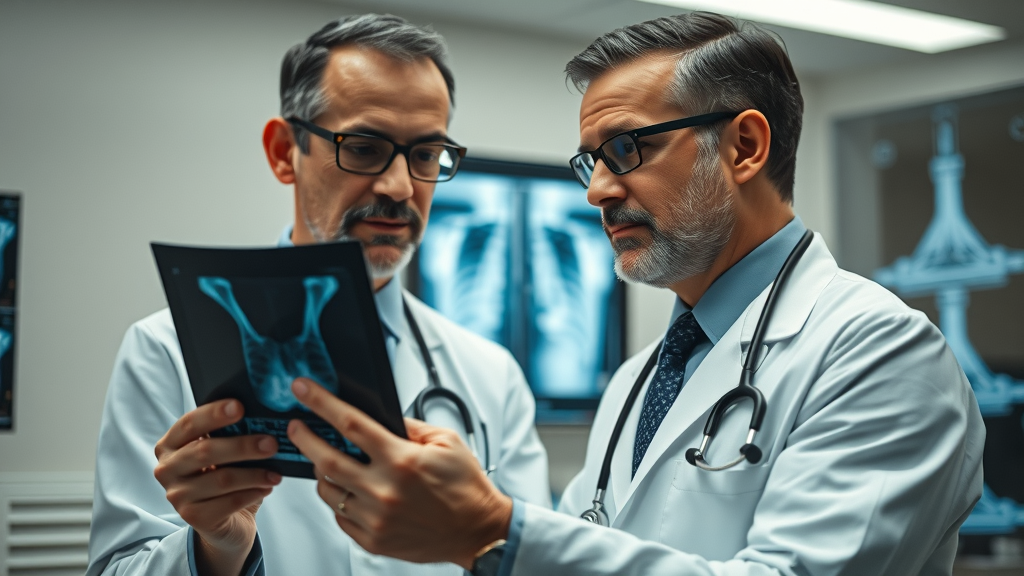
For decades, medical image analysis was limited to the trained eye of a radiologist or specialist who manually inspected X-rays, MRIs, or CT scans. Physicians relied on their expertise and experience to spot anomalies, measure lesions, and provide diagnosis. However, this traditional approach is inherently limited. Human eyesight and cognitive capacity can become overwhelmed by high image volumes or subtle patterns, leading to missed diagnoses or false positives. Furthermore, the sheer complexity and variability of medical images mean that rare or atypical cases can easily be overlooked, even by experts.
With medical imaging growing exponentially, it's nearly impossible for clinicians to analyze every image with the meticulous attention it deserves. Issues like variability between observers and diagnostic fatigue exacerbate the risks. As medical imaging becomes more central to early detection—especially with diseases like breast cancer or stroke—these traditional limitations reveal the pressing need for scalable, automated analysis solutions.
The Advent of Machine Learning and Deep Learning in Medical Imaging
The dawn of machine learning for medical image analysis marked a turning point in healthcare. Advanced deep learning models—especially those based on neural networks—have consistently outperformed traditional image analysis in accuracy and speed. Unlike rule-based or simple statistical methods, machine learning algorithms can rapidly process and learn from vast imaging datasets, identifying complex, hidden patterns beyond human recognition. In recent years, innovations in computer vision and deep learning have enabled automated detection and segmentation of tumors, improved disease classification, and enhanced workflow efficiency for radiologists and clinicians alike.
As these technologies evolve, they're not just supplementing the efforts of healthcare professionals; they're elevating the field to new levels of diagnostic precision. From automatic measurement tools to AI-driven decision support, the integration of machine learning into medical imaging is leading to faster, more reliable, and often life-saving insights.
“Deep learning models now outperform traditional approaches in accuracy, speed, and scalability for complex diagnostic tasks.”
Core Technologies: Key Machine Learning Algorithms Transforming Medical Image Analysis
How Deep Learning and Neural Networks Enable Automated Image Analysis
At the heart of machine learning for medical image analysis are deep learning and neural network algorithms. These models, inspired by the structure of the human brain, autonomously learn to identify features in medical images—from simple edges to complex organ shapes. Convolutional neural networks (CNNs), a type of deep learning architecture, are especially effective for analyzing CT, MRI, or ultrasound scans. Unlike manual feature selection, CNNs extract and prioritize relevant features automatically, enabling them to outperform human-crafted rules in a wide range of diagnostic tasks.
These learning models can be trained on large datasets, improving their ability to spot patterns linked with specific diseases. For instance, an AI trained to recognize diabetic retinopathy can analyze thousands of retinal images, learning to flag microaneurysms or hemorrhages that signal early disease stages. Through repeated training and exposure to annotated data, these algorithms achieve remarkable accuracy and consistency—enhancing rather than replacing the work of radiologists and specialists.
Convolutional Neural Networks: The Backbone of Medical Image Analysis
Convolutional neural networks (CNNs) have become the primary deep learning model utilized in medical image analysis due to their proficiency in handling spatial hierarchies in images. CNNs are specifically designed to analyze pixel relationships and spatial patterns, crucial when assessing high-resolution medical images for anomalies such as tumors, cysts, or lesions. By progressing through multiple layers of automated feature detectors, CNNs localize relevant image regions—normalizing variations in brightness and size—and empower precise image classification and segmentation tasks. Their robustness stems from their adaptability to different types of imaging data, whether grayscale X-rays, 3D MRI scans, or colored pathology slides.
This adaptability allows CNN-based models to excel at both binary (disease/no disease) and multi-class classification, significantly increasing diagnostic throughput. As newer architectures—like ResNet or U-Net—become mainstream in clinical AI, their ability to handle increasingly complex image tasks continues to push the envelope for medical image segmentation, detection, and risk prediction.
Comparing Imaging Data Handling: Machine Learning Algorithms vs. Traditional Computer Vision
Traditional computer vision relies on pre-designed, handcrafted features for analyzing medical images. These rule-based methods are suitable for standardized, well-understood tasks, but they struggle with the variability and subtlety present in real-world imaging data. By contrast, machine learning algorithms, particularly deep learning models, use raw pixel data to uncover patterns and anomalies that would go undetected with classical approaches. This means deep learning is better at scaling, adapting, and maintaining high accuracy across diverse datasets.
Moreover, with machine learning for medical image analysis, the model's capacity to self-learn from annotated datasets eliminates many human-induced biases, enabling more consistent and objective results. While traditional computer vision may offer interpretability and simpler computational needs, its tradeoff is usually lower accuracy and less flexibility for evolving diagnostic challenges.
| Metric | Deep Learning Models | Classical Learning Models |
|---|---|---|
| Accuracy | High (often >97% in disease detection tasks, such as breast cancer diagnosis) | Moderate to High (but lower than deep learning for complex images) |
| Speed | Fast (real-time analysis possible with GPUs) | Slower (manual feature extraction required) |
| Common Use Cases | Automated image segmentation, disease classification, anomaly detection | Simple anomaly detection, image enhancement, basic measurements |
| Scalability | Highly scalable with large datasets and complex tasks | Limited, struggles with large and diverse datasets |
Machine Learning for Medical Image Analysis in Action: Case Studies & Success Stories
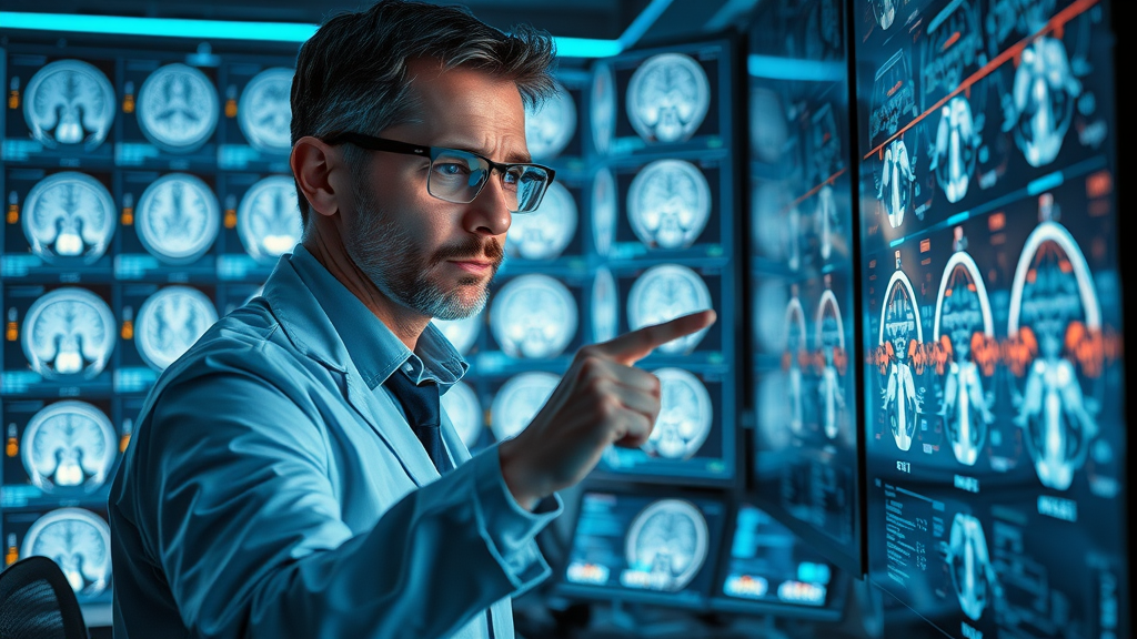
Image Classification for Disease Detection
Machine learning for medical image analysis has achieved spectacular results in disease detection through automated image classification. Instead of relying solely on human eyes, deep learning models correlate imaging patterns—such as tumor shapes, densities, or shading—with thousands of confirmed diagnoses, dramatically improving sensitivity and specificity. For example, algorithms now surpass human radiologists in identifying early-stage lung nodules in CT scans and have set new benchmarks in breast cancer screening. This computer-based approach reduces diagnostic backlog and ensures that vulnerable patients receive attention before diseases progress.
These automated systems also play a critical role in resource-limited settings where access to expert radiologists is restricted, further democratizing access to top-tier medical imaging diagnostics globally.
Semantic Image Segmentation and Tumor Localization
One of the defining strengths of machine learning lies in image segmentation—the process of automatically outlining regions of interest, such as tumors or lesions, on medical images. Semantic segmentation enables not just detection, but precise measurement of abnormal regions, which is crucial for planning treatment and monitoring disease progression. Deep learning models, particularly U-Net and similar convolutional neural networks, have set new standards for accuracy in segmenting complex organs and small pathologies.
By reducing variability in tumor measurement and ensuring consistency across patient scans, these tools provide clinicians with highly reliable data for making treatment decisions and tracking therapy effectiveness over time.
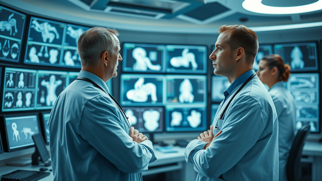
Improving Diagnostic Accuracy in Radiology with Computer Vision and Deep Learning
The fusion of deep learning and computer vision not only accelerates image analysis workflows but also significantly elevates overall diagnostic accuracy. In daily clinical practice, these models support radiologists by flagging high-risk images, prioritizing urgent findings, and minimizing oversight. This technology's integration with PACS (Picture Archiving and Communication Systems) ensures immediate and seamless access to AI-powered analytic insights.
Such advancements empower radiologists to make faster, better-informed decisions, directly impacting patient outcomes, especially in time-sensitive conditions like stroke or cancer metastasis.
- Breast cancer detection using deep learning algorithms
- Lung nodule segmentation with neural networks
- Diabetic retinopathy assessment via automated image analysis
Expert Perspectives: The Promise and Pitfalls of Machine Learning for Medical Image Analysis

“While artificial intelligence accelerates diagnosis, only a multidisciplinary approach ensures clinical safety and ethical considerations are addressed.”
Ethical Dilemmas in Using Artificial Intelligence for Medical Imaging
The rapid expansion of artificial intelligence and machine learning for medical image analysis brings significant ethical challenges. Issues like informed consent, algorithmic transparency, and liability for errors must be front and center in every deployment. For example, when a machine learning model misclassifies a tumor or misses an anomaly, responsibility still lies with human experts—raising critical questions about trust, oversight, and regulatory compliance.
As these learning algorithms move from pilot projects to routine care, continuous collaboration among clinicians, ethicists, and technologists is essential to ensure ethical frameworks keep pace with technological innovation.

Data Quality, Privacy, and Transparency in Deep Learning Models
Data quality stands as the pillar of effective deep learning and machine learning models in healthcare. Models need large, well-annotated, and unbiased imaging datasets to deliver trustworthy results. Furthermore, privacy concerns intensify as more medical images are shared across hospitals or even continents; secure, anonymized data handling is not optional—it’s mandatory. Transparency also matters: clinicians and patients must understand not only what the model predicts but also why. This demands explainable AI and open reporting of algorithm performance, limitations, and edge cases.
Ongoing advancements and regulations such as HIPAA and GDPR play a critical role in ensuring ethical and compliant use of machine learning for medical image analysis.
Overcoming Bias in Machine Learning Training for Medical Images
Bias in machine learning method training can have serious consequences, leading to uneven care or misdiagnosis, especially in underrepresented patient populations. If learning models are trained on datasets lacking diversity, their performance drops for rarer diseases or minority groups. Addressing this means assembling multi-institutional, diverse training datasets and using federated learning, which allows models to learn from decentralized data while preserving privacy. Active monitoring and validation are necessary to minimize and correct algorithmic bias over time, ensuring equitable care for all patients.
Trending Topics: What’s Next for Machine Learning in Medical Image Analysis?
The Expansion of Learning Methods: Federated Learning and Transfer Learning
Next-generation machine learning methods in medical imaging embrace federated learning, a decentralized approach where models are trained across multiple sites without centralizing sensitive patient data. This not only enhances privacy but also broadens the diversity and applicability of learning, improving results for underserved populations. Transfer learning—leveraging pre-trained deep learning models from other domains—drastically reduces the amount of data and time needed to develop new diagnostic algorithms, accelerating clinical adoption.
These techniques pave the way toward more robust, inclusive, and secure models that harness the true variety inherent in global healthcare imaging data.
Towards Explainable Artificial Intelligence for Medical Image Analysis
As deep learning model adoption surges, so does the demand for explainable artificial intelligence (XAI) in medical image analysis. Clinicians want not just a diagnosis, but actionable insights with visual explanations—such as heatmaps showing exactly why a tumor was flagged or which features the model based its conclusion upon. XAI builds clinical trust, supports regulatory review, and empowers experts to verify or question AI decisions, making it indispensable for mainstream deployment.
Continuous research is bridging the gap between AI “black box” models and interpretable, clinician-friendly tools in real-world medical imaging environments.
Integration with Telemedicine and Hospital Workflows
Seamless integration of machine learning into telemedicine platforms and hospital IT systems promises to extend advanced diagnostics far beyond traditional centers. Real-time, AI-driven medical image analysis bolsters point-of-care testing, remote consultations, and secondary opinions, especially in underserved or rural locations. As computer vision and deep learning are embedded in hospital workflows, clinical teams spend less time on repetitive measurements and more on complex, value-driven care, improving the overall patient experience.
Expect hospital systems of the near future to feature collaborative AI dashboards, live alerts, and cross-disciplinary data sharing for a new era in personalized and timely medical imaging diagnostics.
People Also Ask: Answers About Machine Learning for Medical Image Analysis
How does machine learning improve accuracy in medical image analysis?
Machine learning uses advanced algorithms and deep learning models to automatically detect patterns in complex medical images, reducing human error and delivering faster diagnostic outputs.
What are common applications of machine learning in medical imaging?
Typical applications include disease classification (such as cancer), image segmentation for lesion localization, automated measurements, and risk stratification using learning models.
Key Takeaways on Machine Learning for Medical Image Analysis
- Machine learning enhances both the speed and precision of medical image analysis
- Deep learning and computer vision drive major advances in medical imaging diagnostics
- Data integrity and explainability remain crucial as adoption increases
- Future innovations promise even more personalized and real-time diagnostics
FAQs on Machine Learning for Medical Image Analysis
What is the most common machine learning model in medical image analysis?
The most common model is the convolutional neural network (CNN), renowned for its strong performance in image classification and segmentation across modalities like X-ray, CT, and MRI. CNNs can automatically detect and hierarchically process features, making them ideal for diverse medical image analysis tasks.
Can deep learning models replace radiologists?
While deep learning models greatly boost diagnostic accuracy and speed, they are not intended to replace radiologists. Instead, these models serve as powerful decision-support tools, allowing human experts to focus on complex case interpretation, patient communication, and nuanced decision-making that goes beyond what AI can accomplish alone.
How is patient data protected during machine learning analysis?
Patient data is protected using advanced anonymization, encryption, and access controls during machine learning analysis. Regulatory standards like HIPAA and GDPR mandate rigorous data privacy, and emerging techniques like federated learning train models without sharing raw patient images outside hospital networks.
Conclusion: How Machine Learning for Medical Image Analysis is Transforming Healthcare Forever

Machine learning is fundamentally transforming the landscape of medical image analysis, promising a future of faster, more accurate, and accessible diagnostics that empower both providers and patients.
“By embracing machine learning for medical image analysis, healthcare moves closer to a future where diagnostics are faster, more accurate, and accessible to all.”
Take the Next Step with Machine Learning for Medical Image Analysis
Ready to unlock the next generation of healthcare diagnostics? Whether you’re a clinician, researcher, or technologist, learning more about machine learning for medical image analysis is your gateway to revolutionizing medical care. Explore further—innovate boldly and help lead the future of precision medicine!


Write A Comment